Click on this icon to see the image.
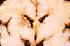
Click on this icon to see a low magnification photo. The lesion is highlighted by the arrow.
NQ-049a Answer: (D) Cavum septum pellucidum
NQ-049b
Answer:
(C) This brain is affected by
periventricular leukomalacia (PVL)
Diagnosis:
Periventricular leukomalacia (PVL)
Pathology of the cases:
·
Age of the specimen:
There is practically no myelin here and judging by the shape of the gyri, this
is a fetal brain and this is, in fact, a brain from a fetus with 30 weeks of
gestation. Myelination of the cerebral hemispheres and the cerebellum is largely
a post-natal event. At the time of birth, there is very little myelin in the
cerebral hemispheres and the cerebellum. However, by the time an toddler has
reached the age of 2 years old, the pattern of myelin is very similar if not
identical to that of an adult on gross examination.
·
Periventricular lesion:
If you look carefully, you will find a subtle chalky irregular lesion at a short
distance from the ventricle (black arrow) surround by tissue that looks like it
is breaking down. There are also some larger chalky lesions (red arrow) that
correspond roughly to the internal capsule. These chalky lesions are all
resulted from calcification due to necrosis of the brain tissue. The second
location (red arrow) is not the classic location of PVL but the first one (black
arrow) is.
·
PVL is often seen in premature babies and occurs most commonly in those that are
born at or after 30 weeks of gestation. It is resulted from increased
susceptibility of the white matter to hypoxic ischemic injury due to the
inherent high metabolic rate of white matter during this stage of development.
For premature babies that are born with more prematurity, such as those born
with less than 28 weeks of gestation, germinal matrix hemorrhage is far more
common than PVL.
[click here to see a case]
·
Histologically, PVL occurs as a well demarcated, small, irregular lesion,
sometime with cavity formation, in the white matter at a short distance from the
ventricle. The affected brain tissue is often mineralized and gives the chalky
appearance. Although these chalky spots are very helpful in identification of
PVL on gross examination, they can be very subtle and minute and requires
careful examination for recognition.
|
|
Click on this icon to see the image. |
|
|
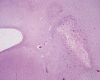
Click on this icon to see a low magnification photo. The lesion is highlighted by the arrow. |
|
|
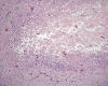
Click on this icon to see the necrotic core and rimming reactive change. |
|
|
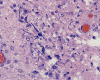
Click on this icon to see the mineralized cells, likely a neuron, highlighted by an arrow. It is these mineralized cells that give the chalky spots. |
Progressive multifocal leukoencephalopathy (PML):
This is usually caused by JC virus in immune compromised patients and is
essentially a disease of adults. The salient features of PML include loss of
myelin with or without the presence of necrosis and atypical glial cells
that contain the virus particles. The viral particle can be demonstrated by
immunohistochemistry, in situ hybridization, and electron microscopy.
Multiple sclerosis (MS): This is not a disease that could be seen in
premature infants or new born simply because it is a demyelinating disease
and by definition there is loss of myelin that is already formed. There is
no myelin to loss at this age group! Multiple sclerosis is rarely seen in
children under 10 years of age and there is a huge female predominance in
this age group. It is uncommon in teenagers. It is common in young adults
and there is also a female predominance.
Kernicterus: This can be seen in this age group and the gross
pathology is characterized by bright yellow discoloration of the cerebral
gray matter. There is no such discoloration here.