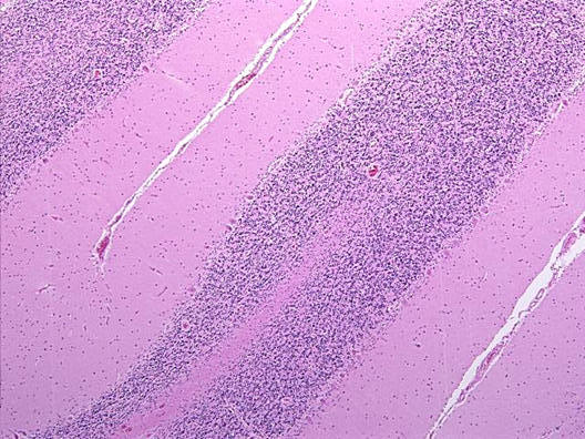
These two images are taken at the same magnification using the same microscope and camera for comparison.
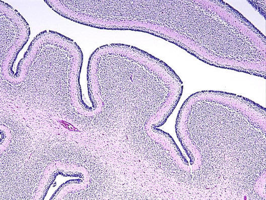
Cerebellar folia, term infant, HE stain: Note that the external granular layer is still present. At this stage, the Purkinje cells is well formed and can be recognized as a layer of cells even at this magnification.