This brain was obtained from a 14 year-old boy who has a confirmed diagnosis of Mitochondrial myopathy, encephalopathy, lactic-acidosis, and stroke like episodes (MELAS). What is most appropriate diagnosis in this case?
A. Meningitis
B. Lamina necrosis of cortex
C. Demyelinating changes
D. Cortical dysplasia
E. Territorial infarct secondary to obstruction of an artery
Answer and Discussion: The answer is (B). The areas with pathologic changes is highlighted by the arrow. There is no exudate or any pathologic changes in the leptomeninges to suspect meningitis. The white matter is in pristine condition with no gross evidence of myelin loss to justify a demyelinating process. Cortical dysplasia may lead to abnormal changes on the cortex. However, the shape and thickness of the cortical ribbon may be abnormal but the ribbon should still look like gray matter on gross examination. Territorial infarct secondary to obstruction of an artery would typically involves both gray and white matter which is not the case in this specimen.
The salient pathologic finding is that of a necrotic cortex adjacent to the white matter (arrow). This is classic for lamina necrosis of the cortical ribbon (gray matter). The patient has a history of MELAS. Large neurons are metabolically very active and needs substantial supply of energy. In mitochondrial diseases, the energy supply is not sufficient for the large neurons. Other neurons and the white matter, however, has lower metabolic rate and are able to survive. This lead to necrosis of a lamina of the cortex as the large neurons are found in lamina III and V in the cortical ribbon.
|
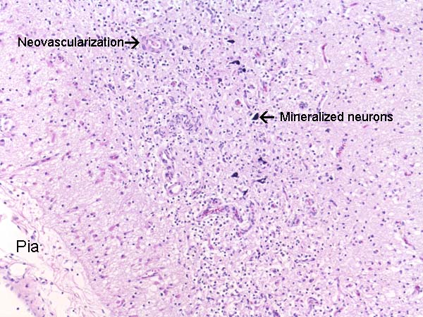
Hematoxylin & eosin |
Histopathology: The layer of lamina necrosis is replaced by mineralized neurons, reactive gliosis and neovascularization. |