Case No.: A-001 Quiz
Diagnosis: Acute Appendicitis
Organ: Vermiform Appendix
Last Updated: 04/23/2017
|
|
|
Hematoxylin & eosin |
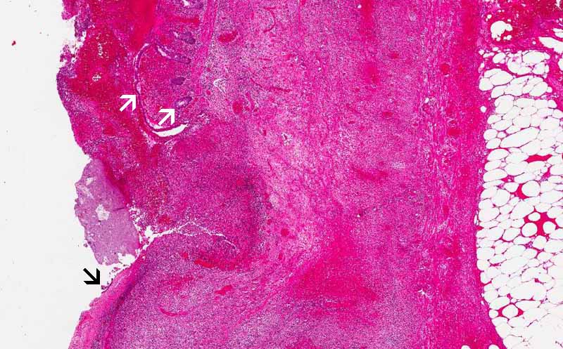
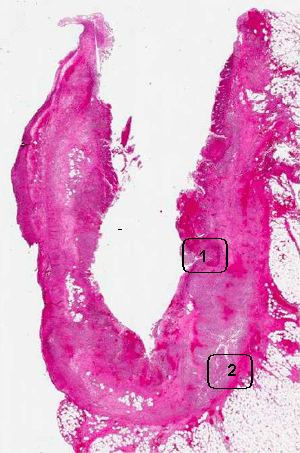
Area 1: This image is taken from the area where there is transmural acute inflammatory cell infiltration that extends from the lumen to the adipose tissue. A large part of the mucosal epithelium is destroyed by the acute inflammation (black arrow). Some residual mucosal epithelium and glands are present (white arrows). |
|
Hematoxylin & eosin |
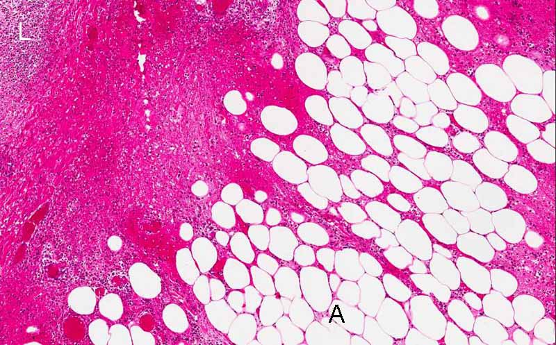
Area 2: The acute inflammation extend through the adipose tissue (A) like a sharp thorn penetraing into a bag of ping pong balls. The lumen side (L) has numerous leukocytes, some are degenerative. |
|
Hematoxylin & eosin |
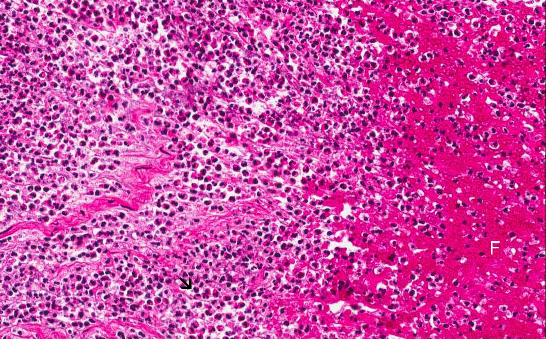
Area 2: The inflammatory cells are almost exclusively neutorphils (arrow) and the inflammatory cells are mixed with fibrin material (F). |
|
History: The patient presented with right lower quadrant pain for three days. An appendectomy was performed and yielded the current specimen.
Histologic Highlights of this Case:
|
Original slide is contributed by Dr. Kar-Ming Fung, University of Oklahoma Health Science Center, Oklahoma, U.S.A.