Case No.: A-002 Quiz
Diagnosis: Diverticulum
Organ: Colon
Last Updated: 08/21/2010
|
|
|
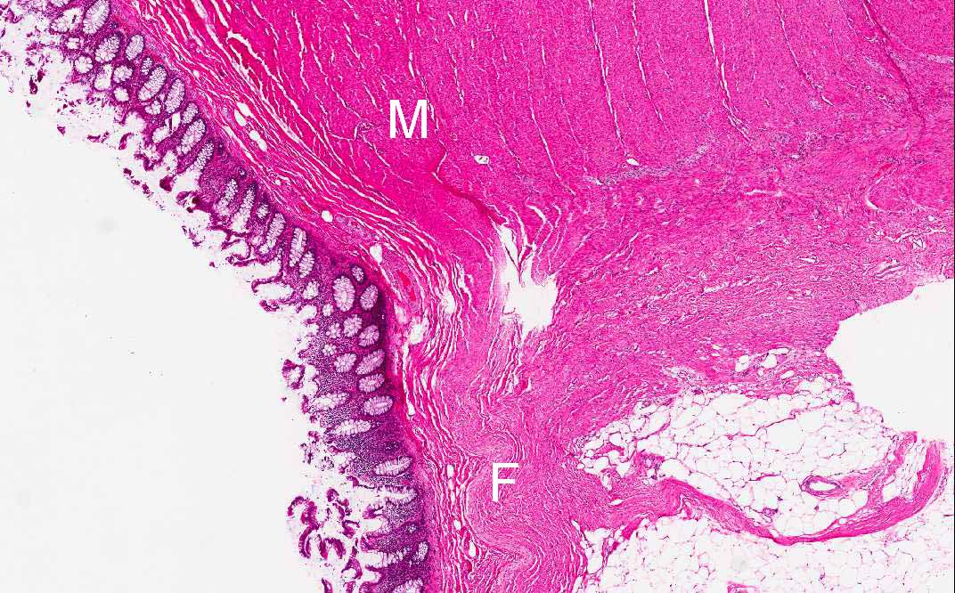
Hematoxylin & eosin |
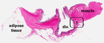
Area 1: This photo is taken at the junction where the diverticulum herniate through the muscle (M). The mechanical support for the flask-shaped structure is only supported by a thin layer of fibrous tissue (F). Note that no inflammatory changes is present in this diverticulum. |
|
History: The patient was a 67 year-old man who had surgery for sigmoid colon cancer. The image being shown here was an incidental finding of the excised colon.
Gross Histologic Highlights of this Case:
Histopathology:
|
Bonus Images:
|
Hematoxylin & eosin |
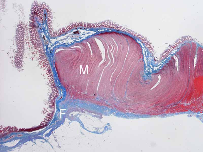
Area 1: This is a Masson' Trichrome stain which stains muscle red and fibrous tissue blue. The relationship between the muscle, the fibrous tissue, and the diverticulum is clearly illustrated here. |
Original slide is contributed by Dr. Kar-Ming Fung, University of Oklahoma Health Sciences Center, Oklahoma, U.S.A.