Online Slide/Full Screen/Open with ImageScope
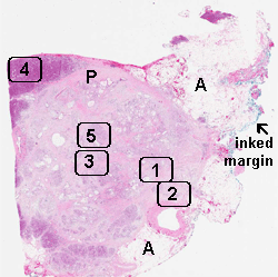
A: Adipose tissue, P: Residual Pancreas
Case No.: A-003
Diagnosis: Invasive ductal carcinoma
Organ: Pancreas
Last Updated: 12/21/2010
|
Online Slide/Full Screen/Open with ImageScope A: Adipose tissue, P: Residual Pancreas |
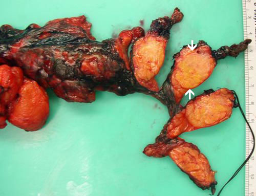 |
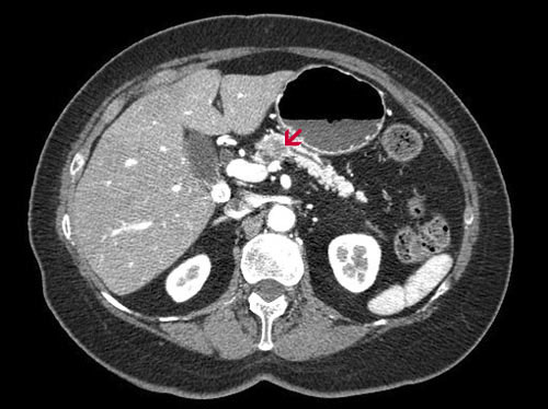
Gross Pathology and CT: The tumor is 1.9 cm in diameter and is relatively well demarcated from the surrounding pancreatic tissue as illustrated here (arrows). It has a lobulated, fibrous cut surface. No gross necrosis or hemorrhage is noted. This relatively well defined mass is reflected in the CT scan. In contrast to most pancreatic tumor, the residual parenchyma of this case is not affected by chronic pancreatitis. |
|
Hematoxylin & eosin |
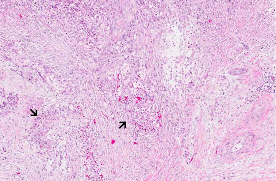
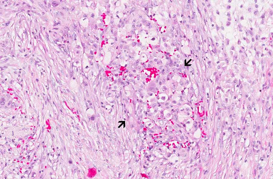
Area 1: The background is very fibrotic and is infiltrated by clusters of neoplastic epithelial cells (adenocarcinoma cells) (arrows). Note that these cells have enlarged nuclei, variation in nuclear size and shpae, and do not have the normal architecture of the normal pancreatic ducts. |
|
Hematoxylin & eosin |
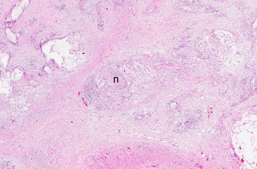
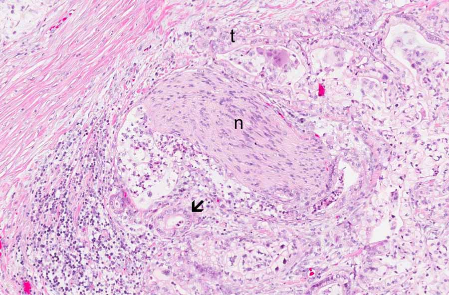
Area 2: The nerve (n) is surrounded and infiltrated by the tumor cells (t). Note that some of the neoplastic cells have lumen formation and this is a genuine feature of adenocarcinoma (arrow). Perineural invasion is a common finding in invasive ductal carcinoma of the pancreas. Note that the tumor cells are mucin producing. |
|
Hematoxylin & eosin |
Area 3: There are large glands with hyperplastic changes and clouding of the epithelial cells accompanied by mild atypia. The level of atypia and architectural changes in these glands are most consistent with pancreatic intraepithelial neoplasm IB (PanIN IB). |
|
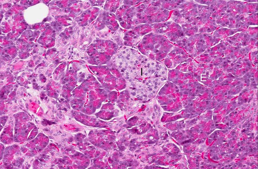
Hematoxylin & eosin |
Area 4: This image is taken at the residual normal pancreatic parenchyma at the rim of the tumor. Note exocrine component (E) and the islets of Langerhans (I). |
|
Hematoxylin & eosin |
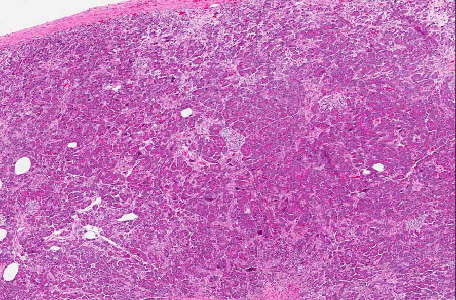
Area 5: Many islets of Langerhans (I) can be found in the fibrotic background. Islets of Langerhans. Islets of Langerhans has a tendency to be preserved within the fibrotic background in pancreatic lesions. They should not be mistaken as tumor cells. |
|
History: The patient was a 75 year-old woman who was presented for sonographic examination of the abdominal for gallstones. A hypoechoic lesion was noted in the pancreas and was later confirmed by CT scan as illustrated in the image here (arrow). As the tumor was in the body of the pancreas, the distal part of the pancreas together with the spleen was excised (distal pancreatectomy) and generated the current specimen.
Histologic Highlights of this Case:
|
Bonus Images:
A endoscopic fine needle aspiration of the lesion was performed and yielded the following images.
|
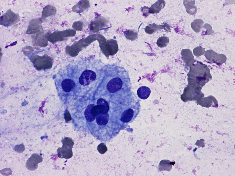
DiffQuick |
Area 1: Carcinoma cells tend to cluster together as illustrated here in this FNA smear. Note that the cytoplasm is rather bubbly appearing due to the mucin content. |
|
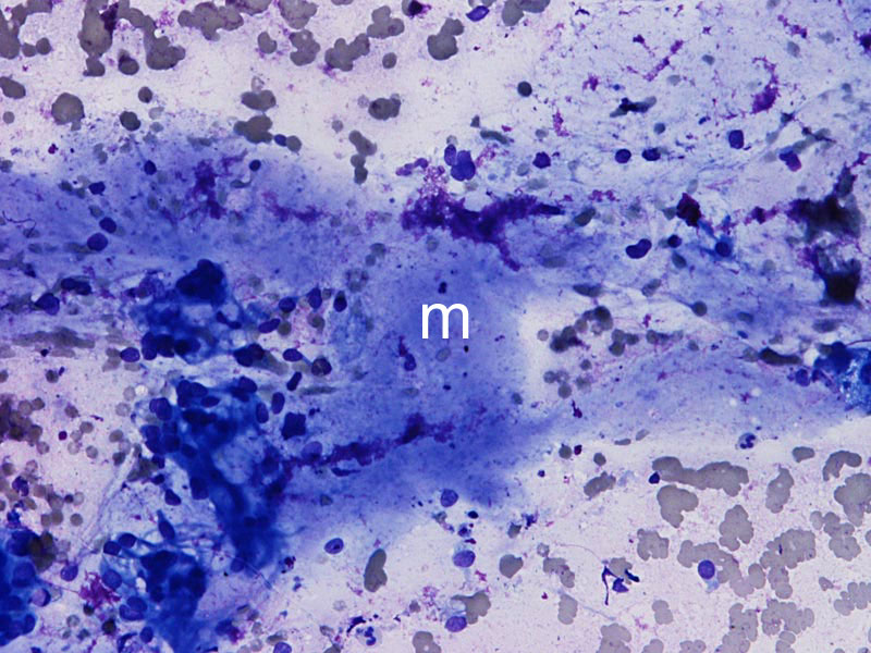
DiffQuick |
Area 2: This tumor is mucin producing. You can see the mucin on this FNA smear (m). |
|
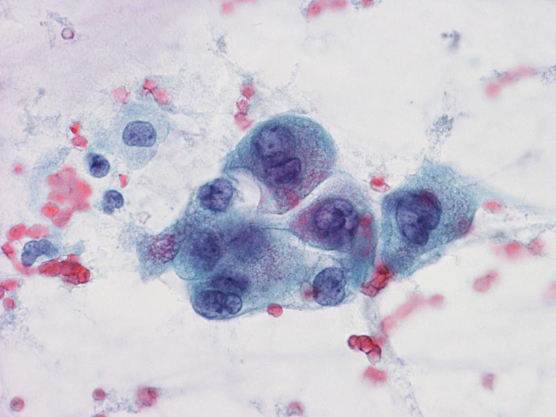
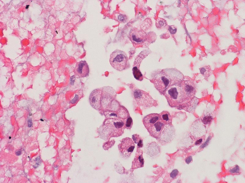
Pap Stain |
Area 3: Nuclear pleomorphism featued by variation in size and shape is well illustrated in this FNA smear. Again, the cytoplasm is bubbly appearing indicating mucin production. |
|
Hematoxylin & eosin |
Area 4: This is an image prepared by sectioning the material obtained at FNA (cell block). Note that the bluish cytoplasmic content corresponds to mucin. |
Original slide is contributed by Dr. Kar-Ming Fung, University of Oklahoma Health Sciences Center, Oklahoma, U.S.A.


