Case No.: A-008
Diagnosis: Periappendiceal abscess
Organ: Appendix
Last Updated: 3/21/2011
|
|
|
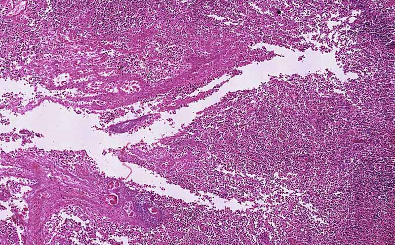
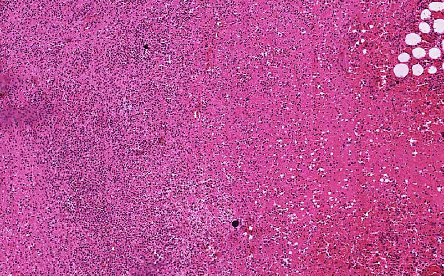
Hematoxylin & eosin |
Area 1: The general outline of the lymphocytes of the normal appendix as well as the shape of the appendiceal lumen can still be recognized. On higher magnification, you can see that many of the cells are not viable. Numerous necrotic cells are present. |
|
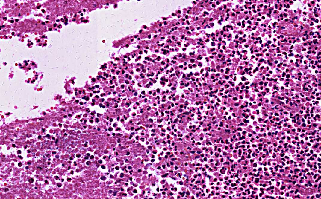
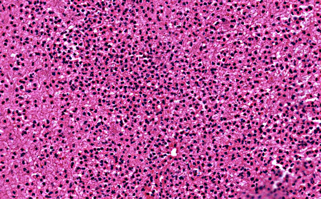
Hematoxylin & eosin |
Area 2: The lumen of the appendiceal wall is necrotic and contains substantial viable or non-viable neutrophils. Congested blood vessels are also present. |
|
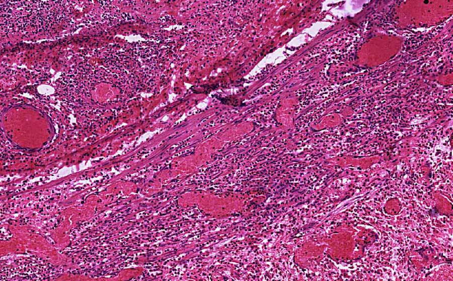
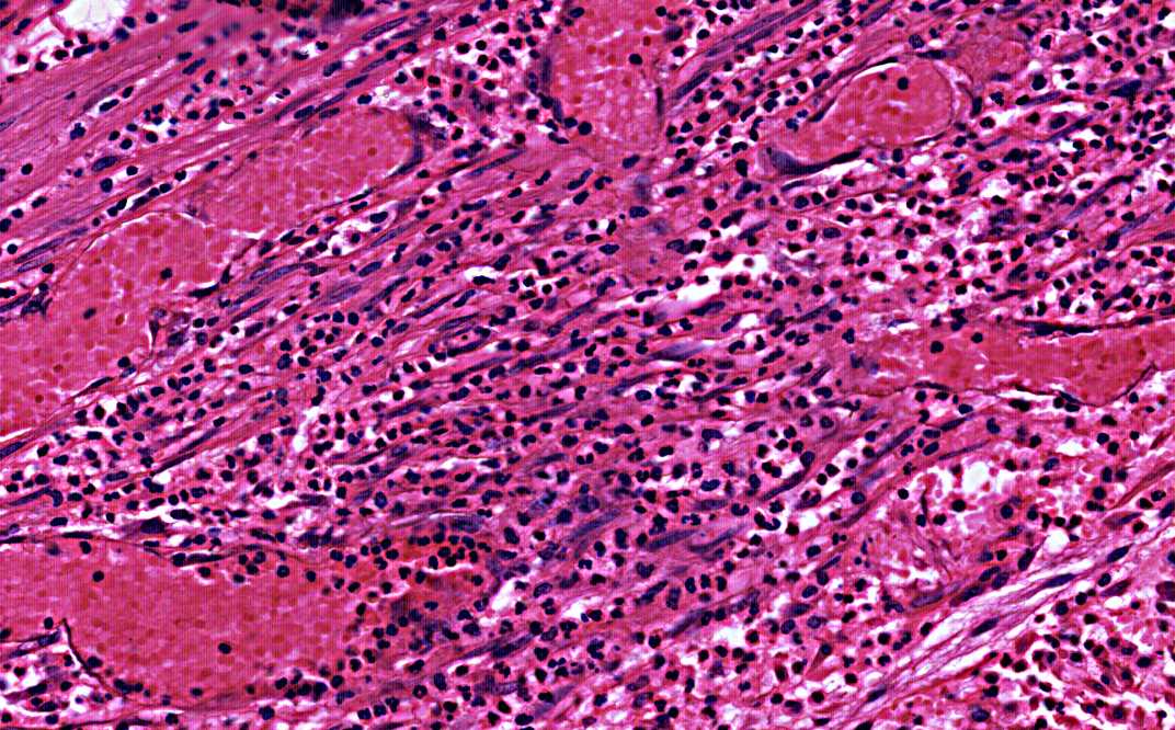
Hematoxylin & eosin |
Area 3: The core of the abscess is essentially a pool of necrotic neutrophils. |
|
History: The history of this case was not know as it was taken from the archival material. However, patients affected by this condition would typically experience right lower quadrant pain with progressive worsening, fever, and chills. In untreated cases, sepsis and death may occur.
Histologic Highlights of this Case:
Comment: Periappendiceal abscess is most often caused byappendicitis with rupture into the periappendiceal tissue and the inflammation is being confined in the periappendiceal tissue. |
Original slide is contributed by Pathology Learning Center, University of Iowa (Iowa Image Collection).