|
History: The history of this
case was not know as it was taken from the archival material. However,
patient affected by this condition often have manifestations of the
gastrointestinal tract including constipation, abnormal bowel movement,
and abdominal pain.
Histologic Highlights of this Case:
-
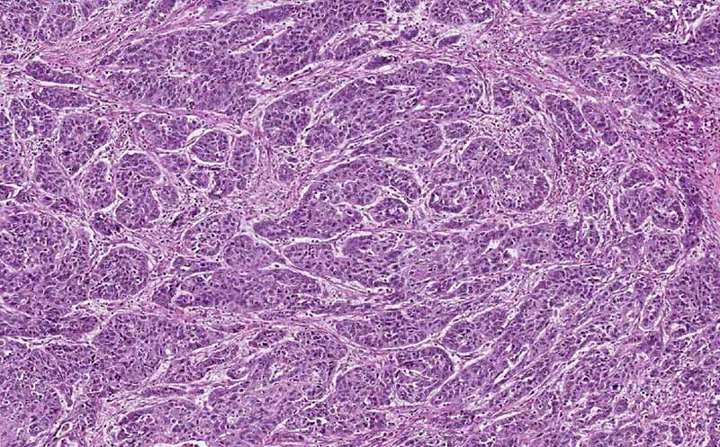
This image is taken from the wall of a
segment of colon. You can recognize the muscle layer. There are two
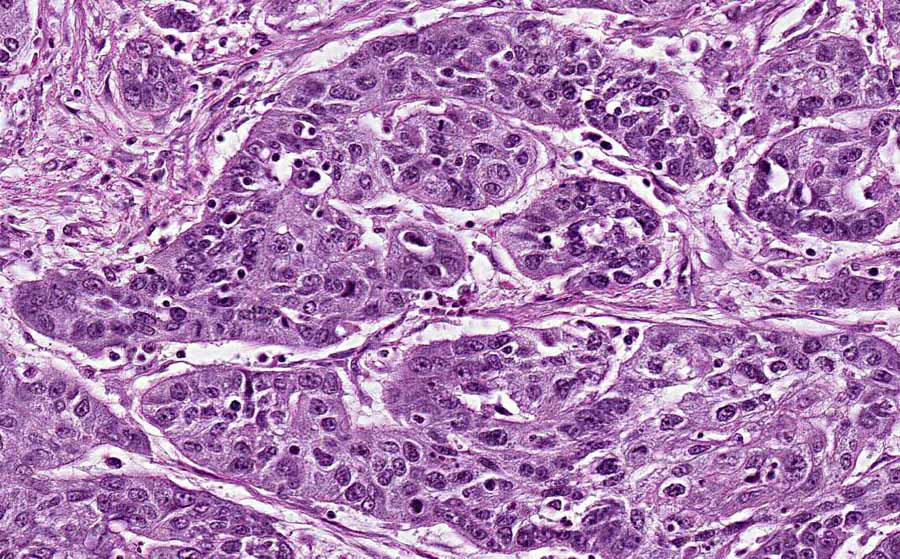
disease processes. The first process is composed of nests of
pleomorphic cells (Area 1). The pathologic features are that of a
neoplasm of the colon. This is most likely the reason why this
segment of colon was surgically removed. The pathology are
suggestive of a carcinoid but no further information is provided in
the archive.
-
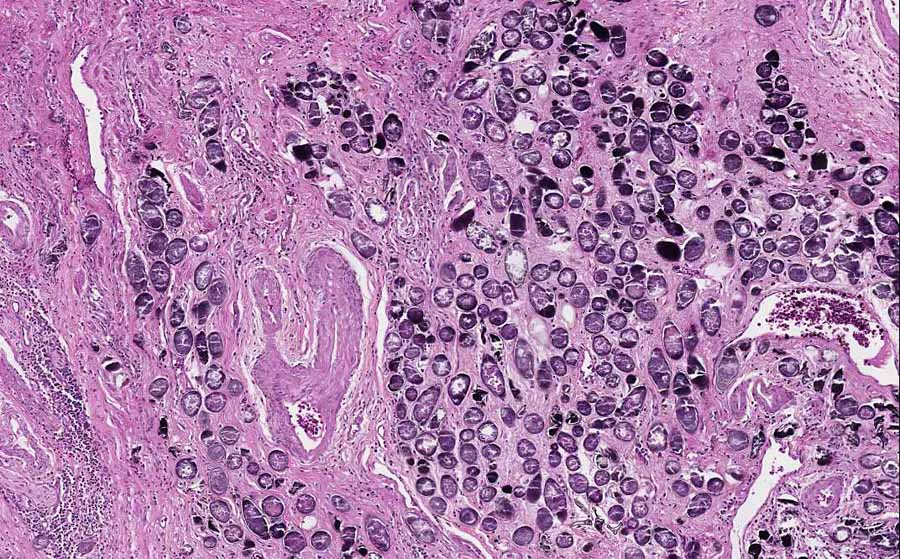
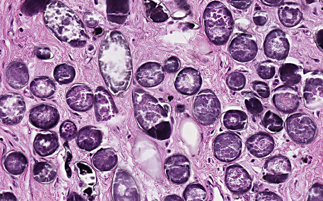
The more interesting lesion, however, is
featured by numerous round to olive shaped structure with a
calcified shell. These organisms are the calcified eggs of
Schistosoma.
Further information:
Schistosomiasis is caused by a trematode,
the Schistosoma. There are 4 species of Schistosoma that can
infect humans. There are some subtle differences in the shape of their
eggs which allow morphological recognition.
- Schistosoma mansoni
and Schistosoma intercalatum: intestinal
schistosomiasis (S. mansoni is found in in South
America and the Caribbean, Africa including Madagascar, and
the Middle East)
- Schistosoma haematobium:
urinary schistosomiasis (S. haematobium found in
Middle East, India, Portugal and Africa)
- Schistosoma japonicum
and Schistosoma mekongi: intestinal schistosomiasis (S.
japonicum and S. mekongi are found in in east and
southeast Asia including China)
|