Case No.: A-012
Diagnosis: Intramucosal adenocarcinoma, moderately differentiated, arising in a background of Barrett's esophagus
Anatomic Region: Gastroesophageal junction
Last Updated: 12/21/2011
|
|
|
Hematoxylin & eosin |
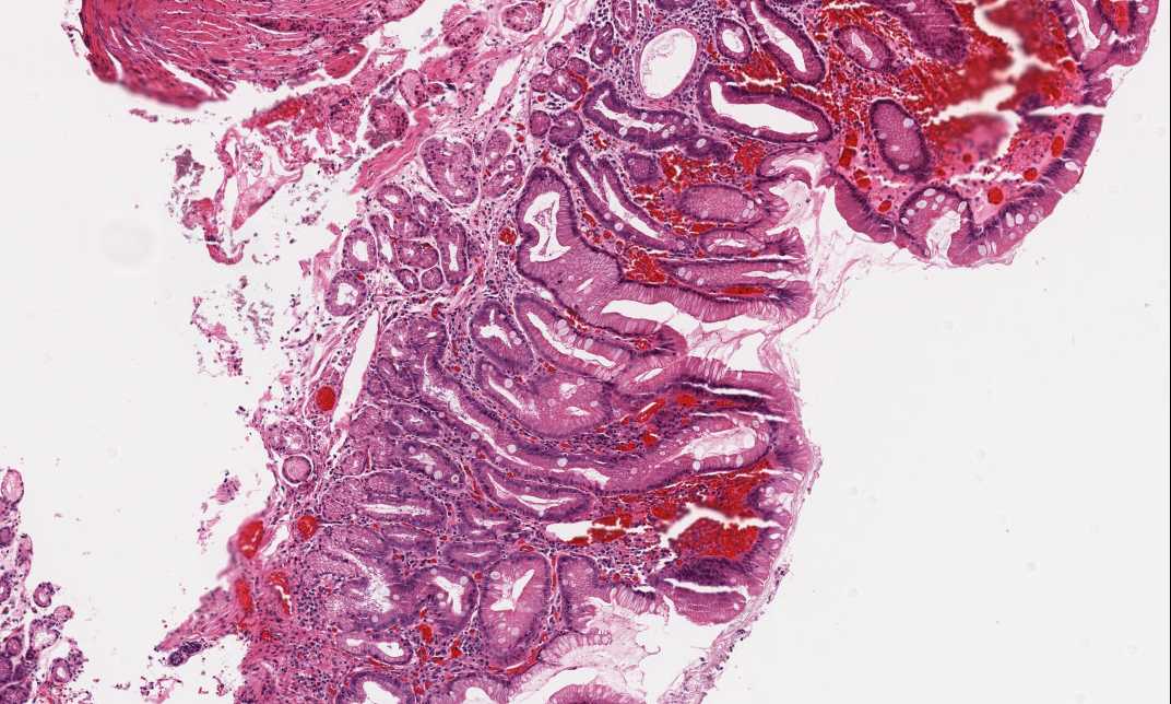
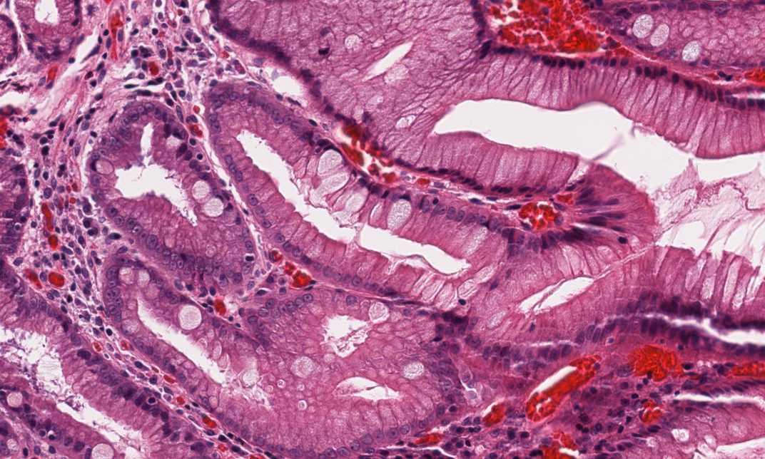
Area 1: In the gastric mucosa, there are some chronic inflammatory cells in the lamina propria. Many goblet cells are present here and they are quite extensive. |
|
Hematoxylin & eosin |
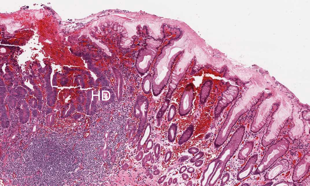
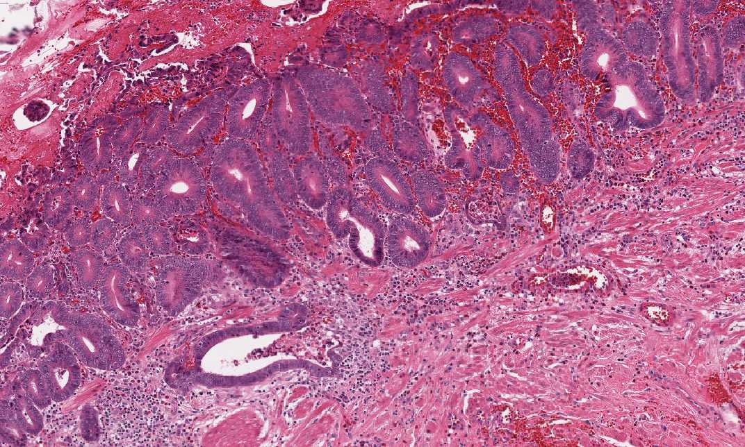
Area 2: Compare the area with high grade dysplasia (HD) and the normal appearing glands on the right of the image. At the interface between the Barrett's esophagus and high grade dysplasia, there are also glands with high grade dysplastic change (HD) and with goblet cell formation. |
|
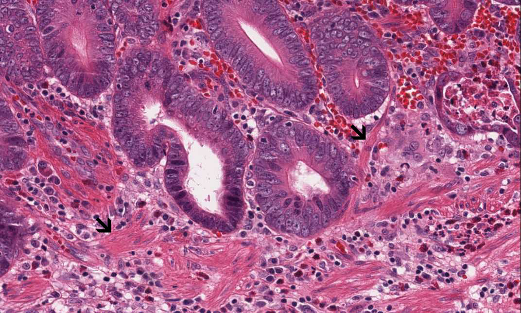
Hematoxylin & eosin |
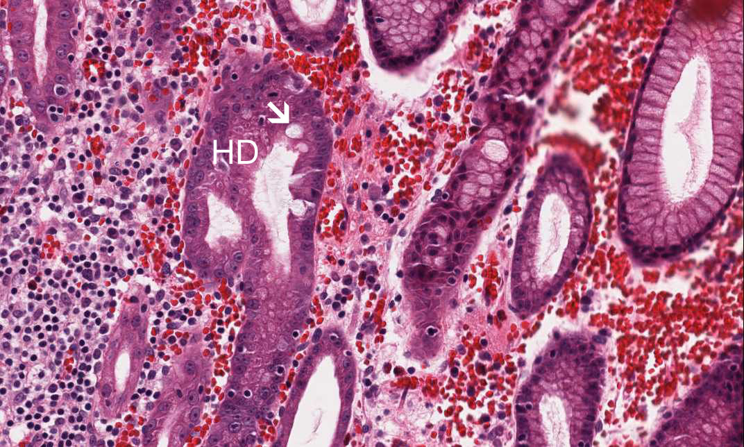
Area 3: The muscularis mucosa is not penetrated as illustrated. However, the tumor cells has already reached the smooth muscle fibers (arrow). |
|
History: This is a mucosal resection from the gastroesophageal junction of an adult. What is you diagnosis?
Histologic Highlights of this Case:
|
Original slide is contributed by Dr. Kar-Ming Fung, University of Oklahoma Health Science Center, Oklahoma, U.S.A.