Case No.: A-014
Diagnosis: Adenocarcinoma, low-grade, invading submucosa
Organ: Colon
Last Updated: 12/21/2011
|
|
|
Hematoxylin & eosin |
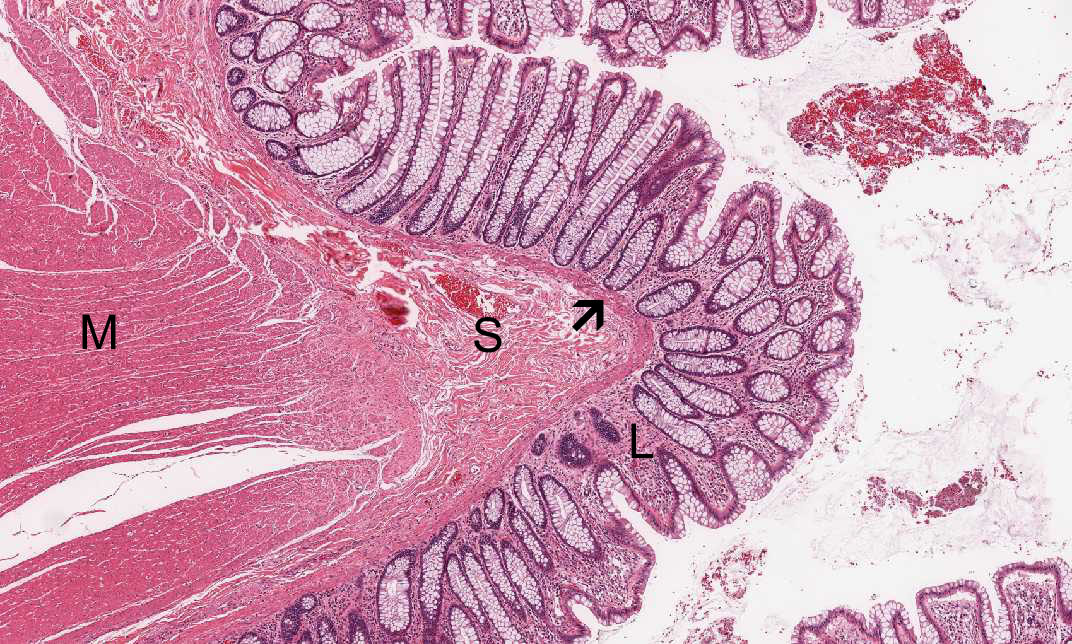
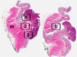
Area 1 (normal area for comparison): This is the normal area for comparison. You can see the muscularis propria (M), submucosa (S), muscularis mucosa (arrow), and lamina propria. In this case, you can see that the tumor has invaded through the muscularis mucosa into the submucosa but has not yet invaded into the muscularis propria. The epithelial cells are in orderly single layered columnar arrangement with small, basally located nuclei and mucin production. |
|
Hematoxylin & eosin |
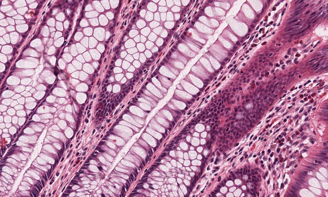
Area 2: In this area, there is area with severe dysplasia (SD). The overall arrangement still follows the normal glands iterms of the relationship between the epithelial cells and the supporting fibrous stroma. Howeve, the epithelial cells have basophilic cytoplasm and no mucin production. The most important part is that the nuclei are much larger and they lost polarity. Many of the nuclei are superficially located. Nest of these abnormal cells have invaded into through the muscularis mucosa into the lamina propria. |
 Hematoxylin & eosin |
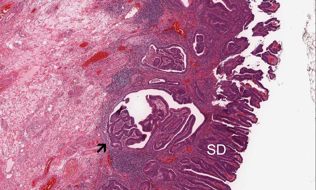
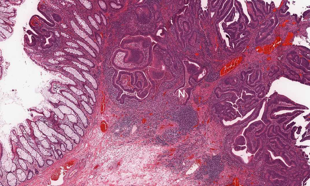
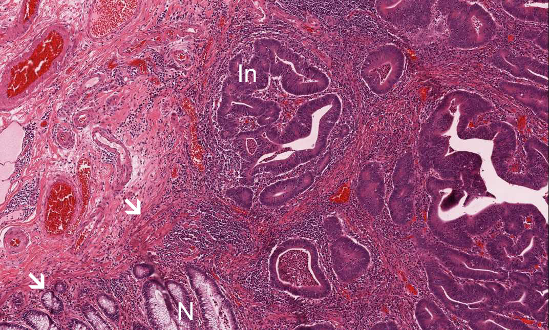
Area 3: This image is taken at the edge of the tumor where you can see the the relationship between the normal area (N), muscularis mucosa (arrow) and the invading tumor (In). |
|
Hematoxylin & eosin |
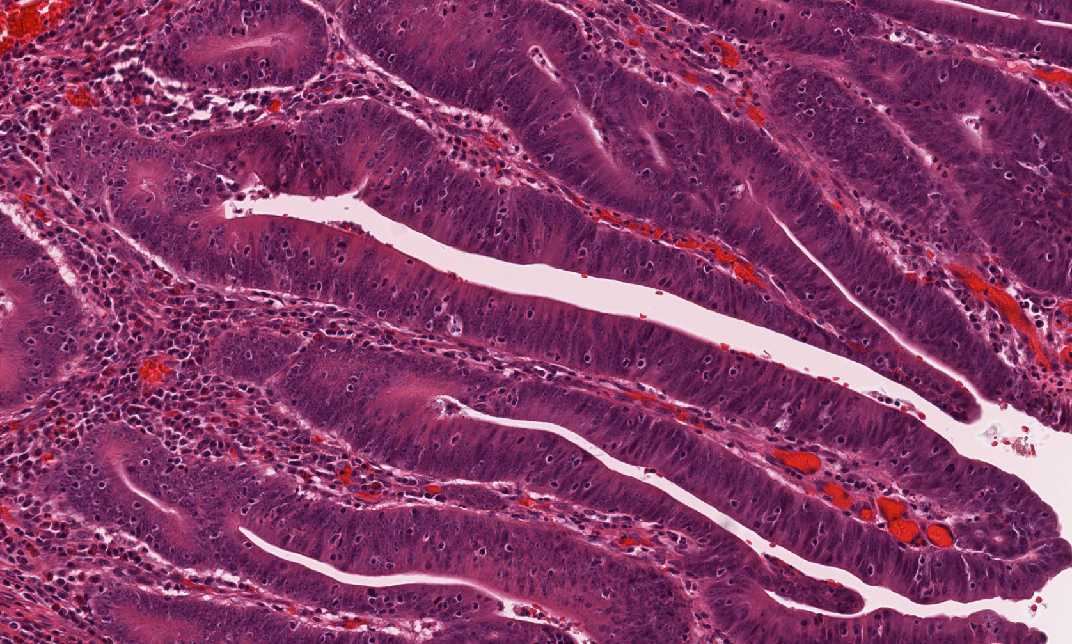
Area 3: This is another area showing the invading carcinoma. Note that necrotic debris are present in the lumens of the neoplstic glands. This is a very common feature for adenocarcinoma of the colon. |
|
History: This specimen was obtained from a colonic resection from an adult. What is your diagnosis?
Histologic Highlights of this Case:
|
Original slide is contributed by Dr. Kar-Ming Fung, University of Oklahoma Health Science Center, Oklahoma, U.S.A.