Diagnosis: Fibroadenoma
Organ: Breast
Last Updated: 12/30/2009
|
|
|
Hematoxylin & eosin |
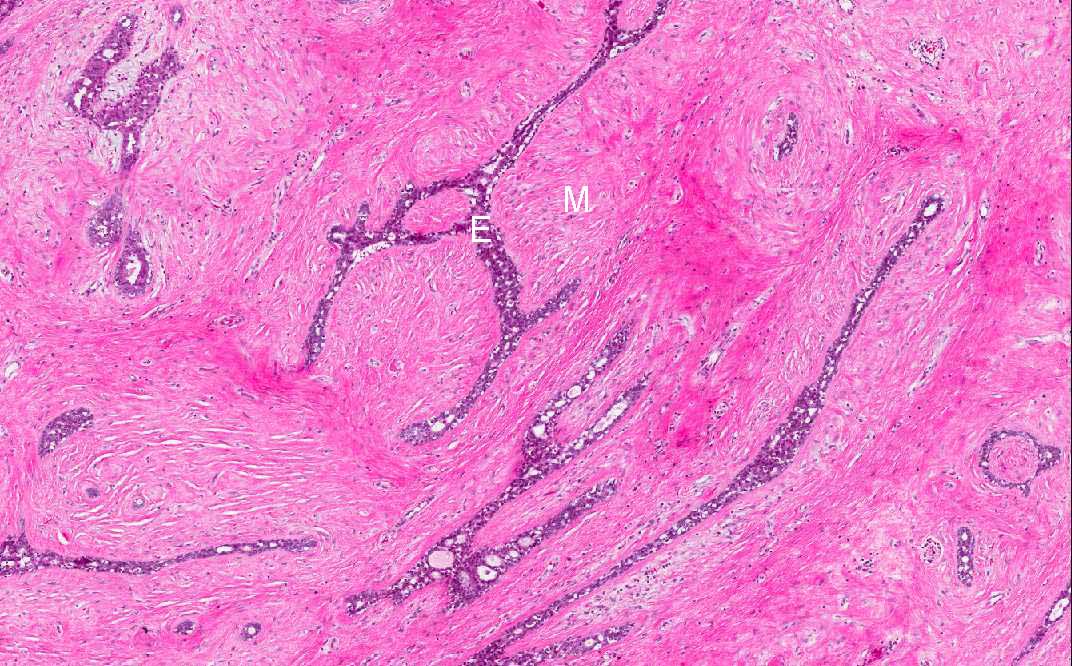
Area 1: In this area you can see both the epithelial component (E) and the mesenchymal component (M). The epithelial component lacks nuclear features indicative of malignancy. The mesenchymal component has low cellularity and with bland nuclei free of enlargement, pleomorphism, prominent nucleoli, or hyperchromatism. |
|
Hematoxylin & eosin |
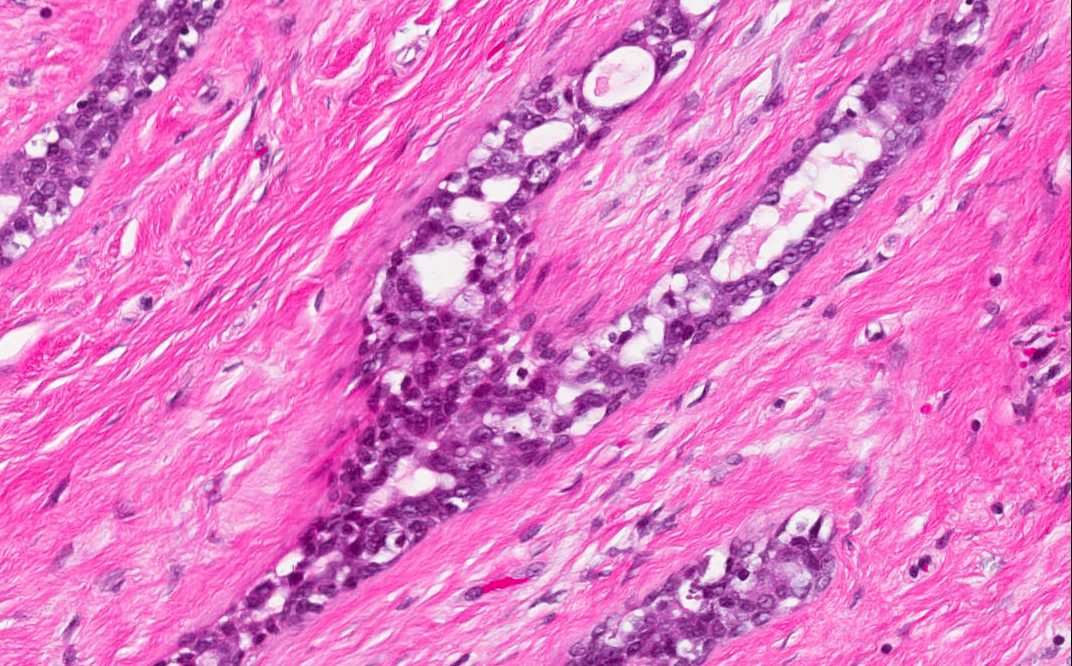
Area 1: Epithelial component does not show any features suggestive of dysplasia or malignancy. The epithelium remains single cell layer. |
|
Hematoxylin & eosin |
Area 1: Low cellularity and fibrotic change in the mesenchymal component. |
|
History: The patient was a 46 year-old woman with a nodule in her left breast. The nodule was firm and movable. This nodule was excised and yielded the current specimen.
Histologic Highlights of this Case:
|
Original slide is contributed by Dr. Kar-Ming Fung, University of Oklahoma Health Sciences Center, Oklahoma, U.S.A.