Case No.: B-003

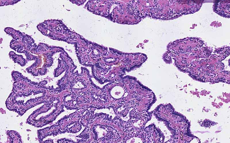
Diagnosis: Intraductal papilloma

Organ: Breast

Last Updated: 3/21/2011
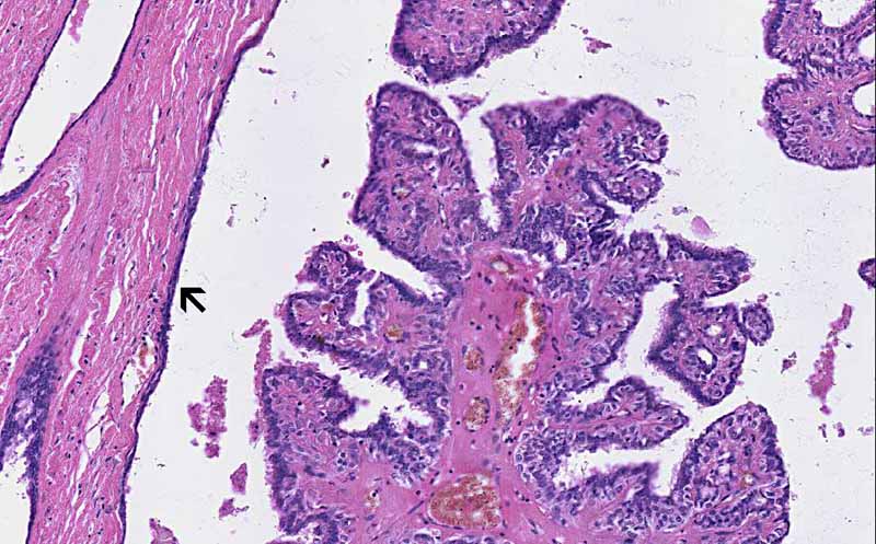
Online Slide/Full Screen/Open with ImageScope

Hematoxylin & eosin

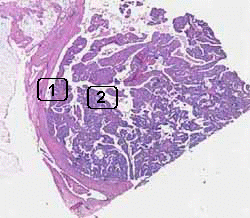
Area 1: The papilloma has dilated the duct. The dilated duct is lined by attenuated epithelial cells (arrow).
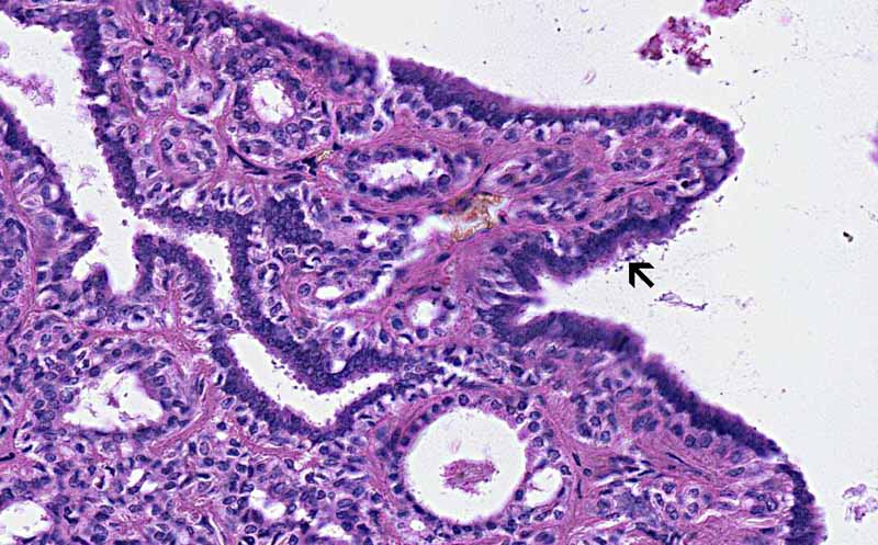
Hematoxylin & eosin

Area 2: The lining epithelial cells (arrow) are single layered, without nuclear atypia, and do not breach the basement membrane. These are features of benign neoplastic proliferation.

History: This slide was taken from the archive and no history was provided. However, women with this condition often complain of nipple discharge.


Histologic Highlights of this Case:

  • The overall architecture is that of a papillary proliferation within a dilated cystic structure. The wall of the cyst is lined by attenuated epithelial cells (Area 1) which suggest that this is originally very likely to be a ductal structure.

  • The papillary proliferation is composed of fronds of fibrovascular stroma covered by a single layer of epithelial cells without atypical nuclear changes or epithelial hyperplasia (piling up of layers of epithelial cells). The the epithelial cells have not breached the basement membrane and invade into the stroma. (Area 1 and 2)

Comment: 

  • Intraductal papillomas often arise in the laciferous ducts in subareolar and central part of the breast. As illustrated here, the duct is dilated by the papilloma and may be dilated to a cystic extent. The cyst may contain clear fluid, bloody fluid, or even coagulated blood.

Original slide is contributed by Pathology Learning Center, University of Iowa (Iowa Image Collection).

 Home Page