History: This slide was taken from the archive and the history was not provided. This condition, however, typically occur in young women and associated with pregnancy or breast feeding.
Histologic Highlights of this Case:
-
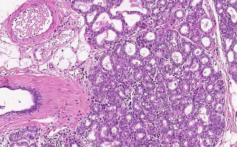
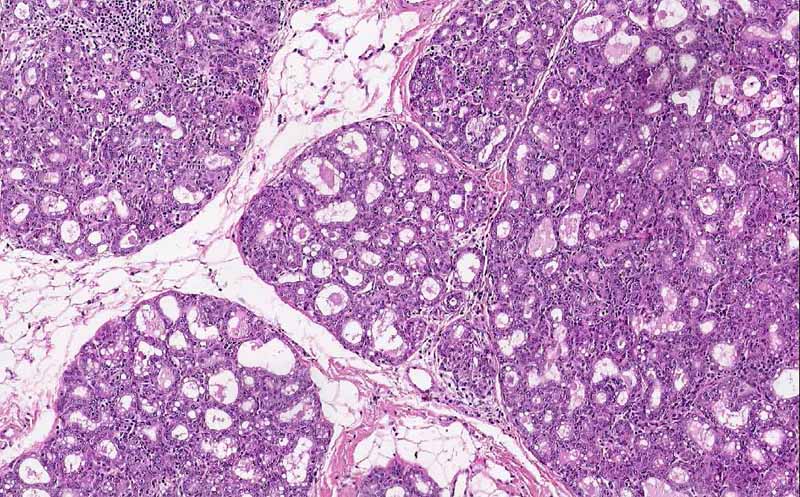
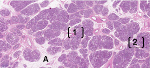
There is proliferation of glands that arrange in lobules and the overall architecture gives a lobular appearance. In many places, the lobules are separated by adipose tissue (A).
-
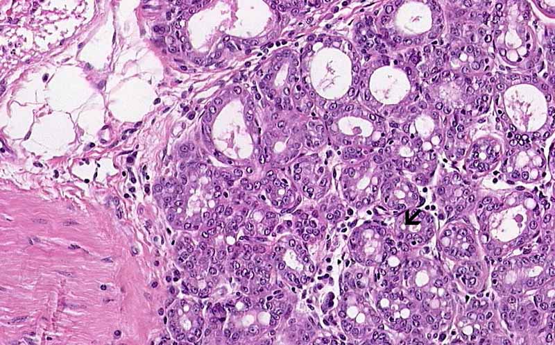
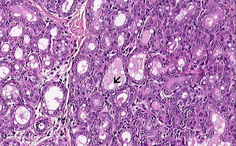
On higher magnification, the glands have a moderate variation in size and some of them are distended. Vacuoles are seen in the content as well as in the cytoplasm of the cells. These vacuoles represent fat of the milk (Area 1 and 2).
-
There is no hyperplastic changes of the epithelium or nuclear atypia.
-
The overall changes are indicative of lactating changes.
Comment:
-
Secretory changes usually occur unevenly throughout the breast during pregnancy. The glandular expansion is accompanied by reduction in fibrofatty stroma. In some extreme cases, most often in the third trimester of pregnancy, these process can form a mass. In the extreme cases, the lobules are separated by only thin fibrous septa and these lesions are known as lactional adenomas.
-
The overall lobular architecture, the tale telling lipid vacuoles, the lack of multiple layers of epithelium (hyperplastic changes) and the lack of nuclear atypia distinguish them from ductal hyperplasia and carcinoma in situ.