Case No.: B-005
Diagnosis: Intraductal papilloma
Organ: Breast
Last Updated: 11/21/2011
|
|
|
Hematoxylin & eosin |
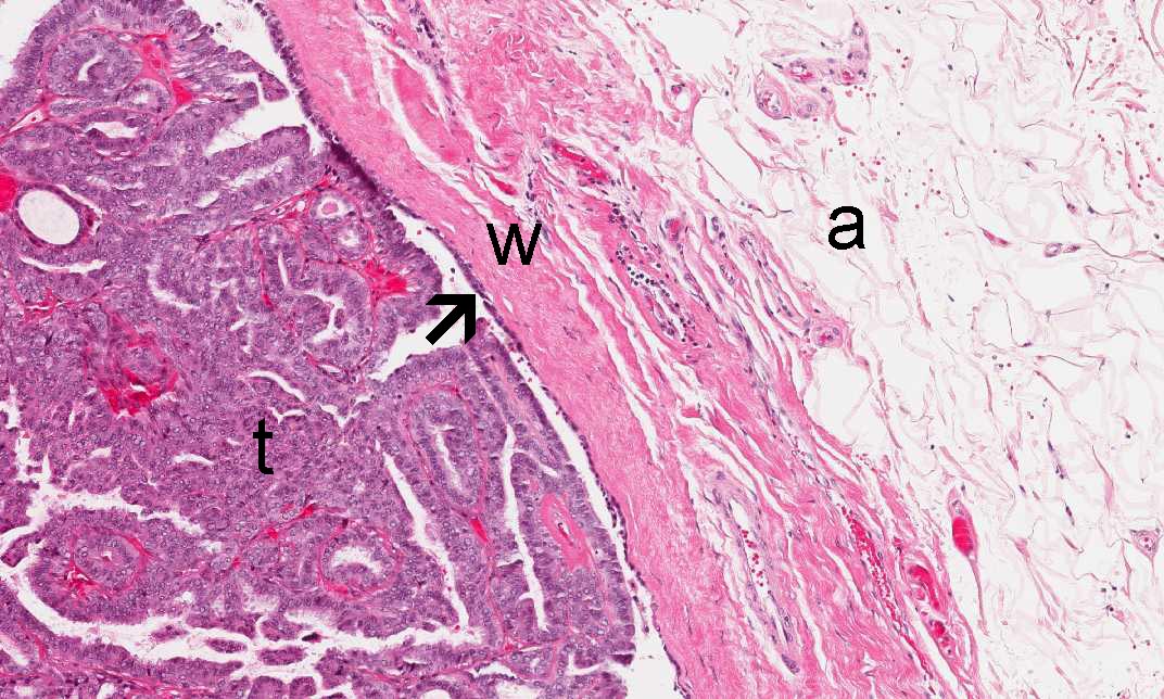
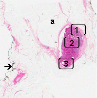
Area 2: Note collagenous wall of the duct (w) and the surrounding adipose tissue (a). The inner surface of the duce is lined by epithelial cells (arrow). Note that there is some hyperplastic proliferation of epithelial cells in this area (t). |
|
Hematoxylin & eosin |
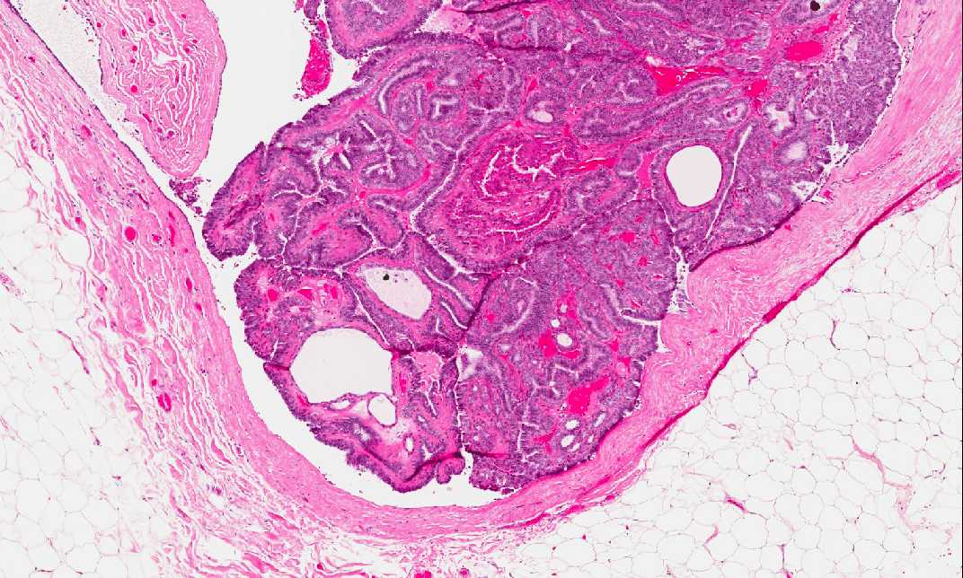
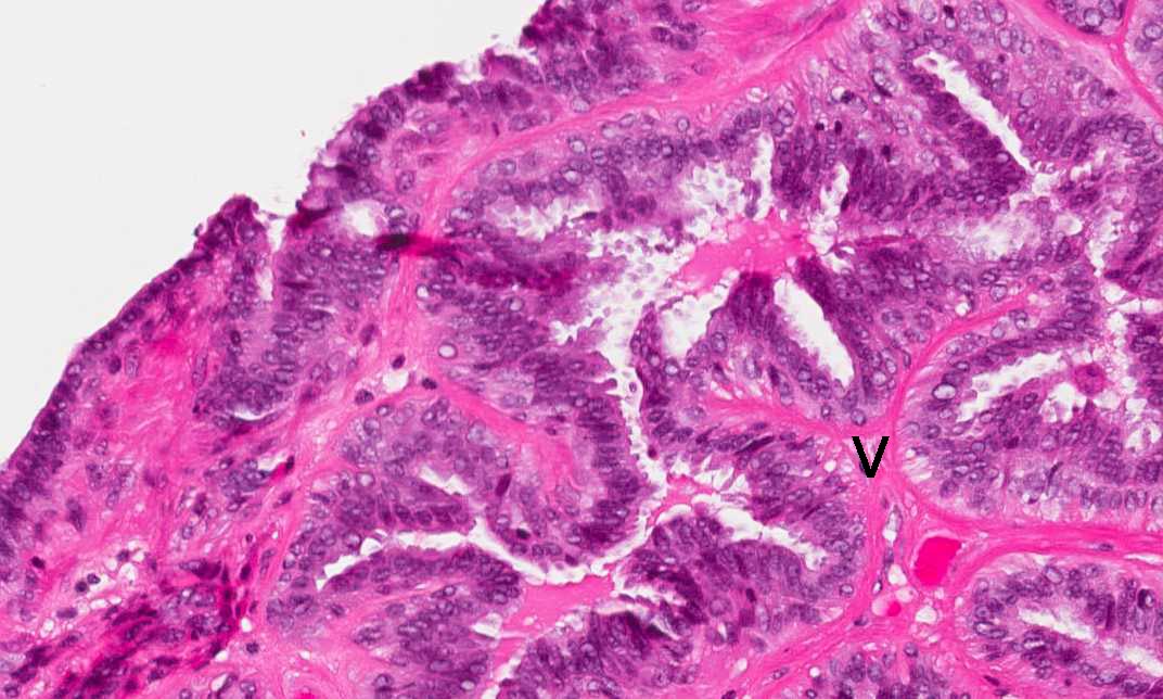
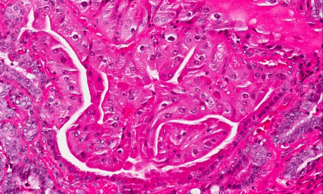
Area 2: Note the papillary, finger-like fibrovascular core (v) that that supports the epithelial cells. There is only one single layer of lining epithelial cells. |
|
Hematoxylin & eosin |
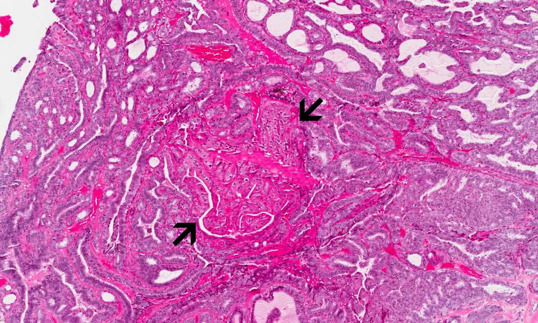
Area 3: Note that there is an island of epithelial cells with slightly increase in size and significantly more eosinophilic than the surrounding cells (outlined by the arrows). These are cells with apocrine metaplasia (oncocytic changes) and is bengin. |
|
History: This slide was obtained from a woman as a mastectomy specimen performed for invasive ductal carcinoma.
Histologic Highlights of this Case:
|
Original slide is contributed by Dr. Kar-Ming Fung, University of Oklahoma Health Science Center, Oklahoma, U.S.A.