Case No.: B-007

Diagnosis: High-grade ductal carcinoma in situ with comedo type necrosis and poorly differentiated invasive ductal carcinoma

Organ: Breast

Last Updated: 1/21/2011
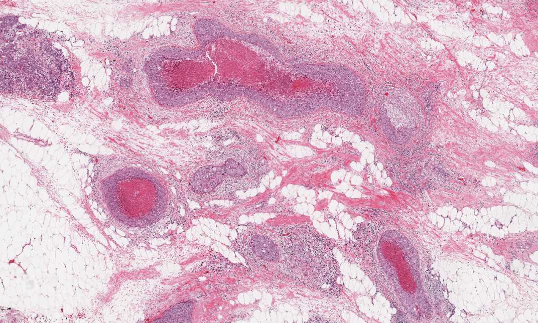
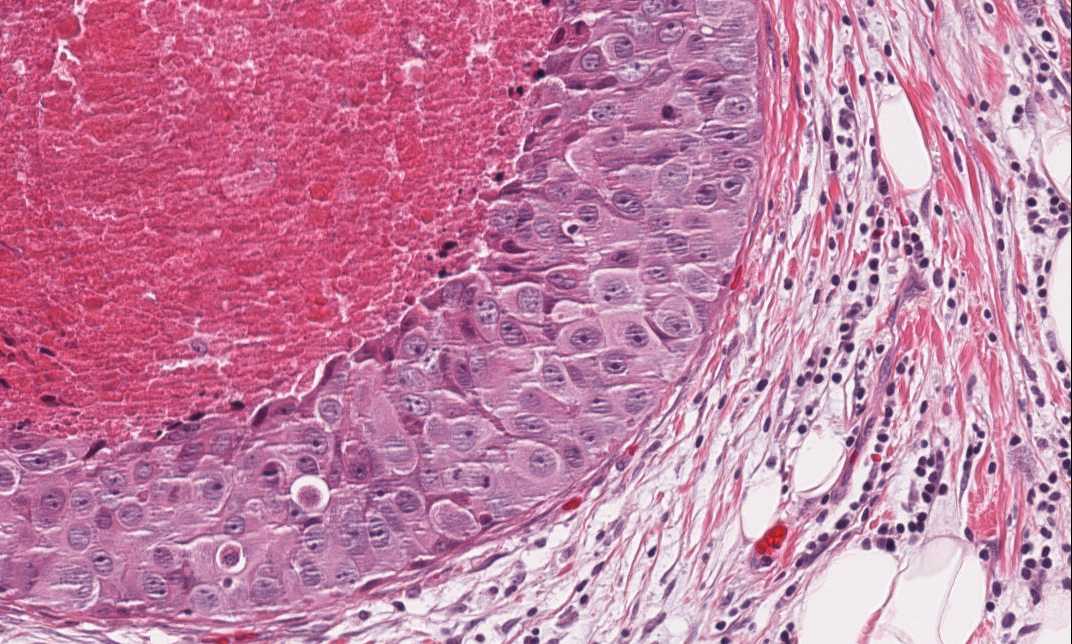
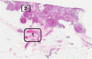
Online Slide/ Full Screen/ Open with ImageScope

Hematoxylin & eosin

Area 1: Ductal carcinoma in situ (DCIS) component.
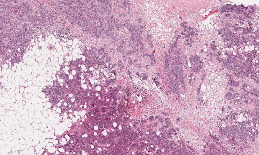
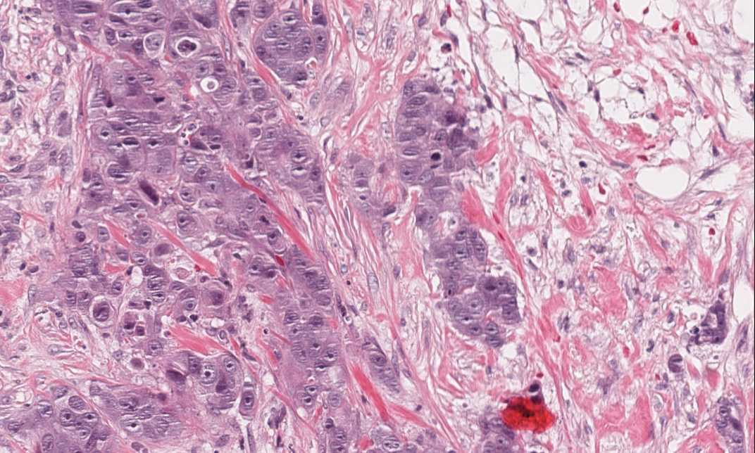
Hematoxylin & eosin

Area 2: Invasive ductal carcinoma component.

History: This specimen was obtained from the breast of a 63 year-old woman. What is your diagnosis?


Histologic Highlights of this Case:

  • In this specimen, both ductal carcinoma in situ (DCIS) and invasive ductal carcinoma are present. The DCIS (Area 1) is composed of large neoplastic cells with large nuclei and prominent nucleoli. These islands have not breeched the basement membrane and the tumor is therefore still in situ.  The coresof these islands are filled with eosinophilic necortic debris (comedo type necrosis). The necrosis seems to have expanded the ducts.

  • The invasive component (Area 2) is composed of invasive cell nests without definitive lumen formation. The tumor cell nuclei are also high grade. These cells are large, with high nuclear to cytoplasmic ratio, pleomorphic, and have distinct nucleoli.

  • Compare this case with a case of well differentiated invasive ductal carcinoma and invasive signet ring cell lobular carcinoma.

Original slide is contributed by Dr. Kar-Ming Fung, University of Oklahoma Health Science Center, Oklahoma, U.S.A.

 Home Page