Case No.: C-005

Diagnosis: Myocardial infarction, acute

Organ: Heart

Last Updated: 3/21/2011

Online Slide/Full Screen/Open with ImageScope

Hematoxylin & eosin

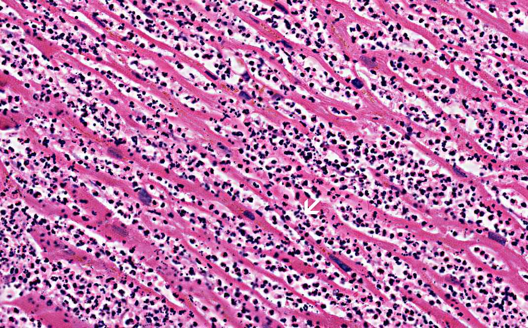
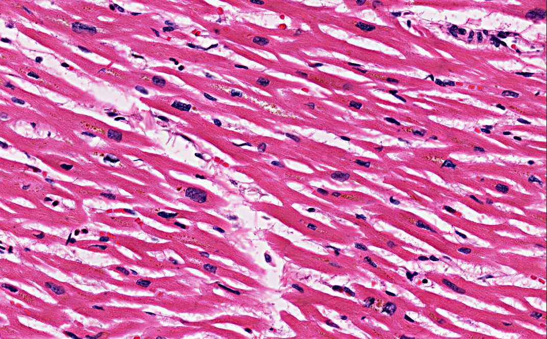
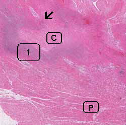
Area 1: Note the intense infiltration of neutrophils (arrow). Can you recognize the residual cardiac muscle fiber in the background?
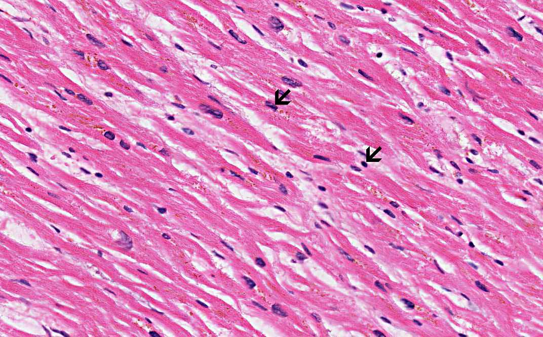
Hematoxylin & eosin

Area C: Note that many of the nuclei are very dark in color (hyperchromatic), small, and have loss nuclear details (arrows).
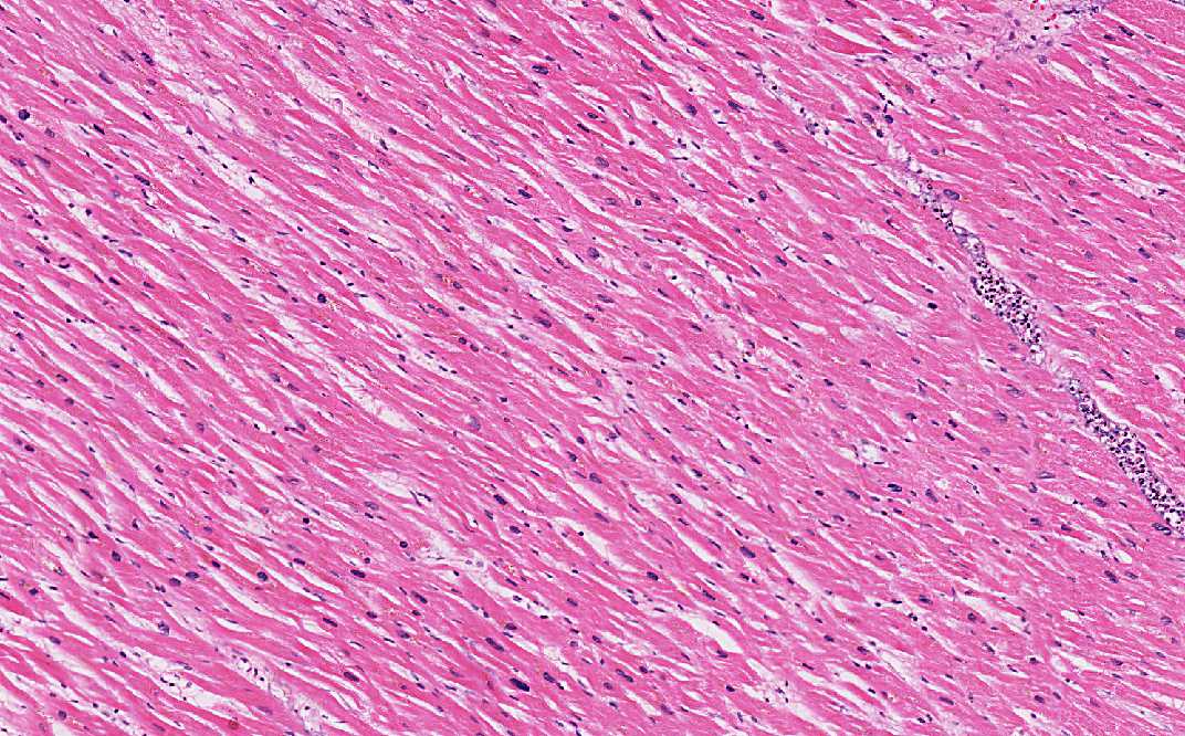
Hematoxylin & eosin

Area P: Compare this area with Area C. The histologic details here are well preserved.

History: This slide was taken from the archive and the history was not certain but it is very likely that the patient had sustained a cardiac emergency a few days before.


Histologic Highlights of this Case:

  • In this autopsy specimen obtained from the myocardium, there are curvy bands of cellular infiltration (arrow) surrounding an imaginary center (Area C). On higher magnification, the cellular areas are intensely infiltrated by numerous neutrophils. This is a case of myocardiac infarction and is likely to be about 3-7 days old as this would be the time that you would see intense infiltration by neutrophils (Area 1).

  • Compare the cardiac fiber from the core of the lesion (C) to those at the periphery (P), you will realize that the core looks more pale in staining (best appreciation at this panoramic view). In addition, the nuclei are hyperchromatic and loss nuclear details. The cell contour is contracted and the striations are lost. These are features of coagulative necrosis. The neutrophils is infiltrating from the outside and eventually will involve the entire lesion should the patient have survived this attack.

Comment:

  • The histopathologic changes are most consistent with an acute myocardiac infarction that would be at the most a few days old (about 3 days). At this stage, there is intense neutrophilic infiltration and coagulation necrosis. Macrophages has not started infiltrating and fibroblastic proliferation has not started either. At this point, the muscle is most fragile and most likely to have rupture. Once when fibroblastic proliferation starts, the tissue will become increasingly fibrotic and less likely to rupture.

Compare this with other stages of myocardial infarction: C006, C007

Original slide is contributed by Pathology Learning Center, University of Iowa (Iowa Image Collection).

 Home Page