Case No.: C-010
Diagnosis: Organizing thrombi associated with atrial fibrillation
Organ: Heart, atrium
Last Updated: 11/21/2011
|
|
|
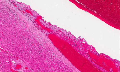
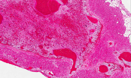
Hematoxylin & eosin |
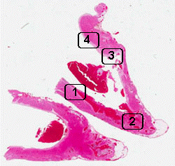
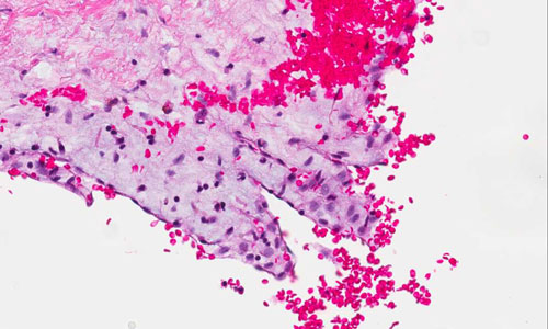
Area 1: These images are obtained at the edge of the thrombus where many foamy macrophages are present. |
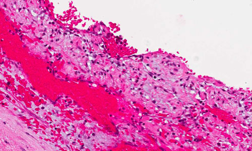
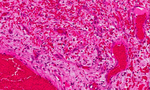 Hematoxylin & eosin |
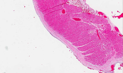
Area 2: These images are obtained at the deep part of the thrombus where many fibroblasts admixed with foamy macrophages are present. The fibroblasts appears spindly. |
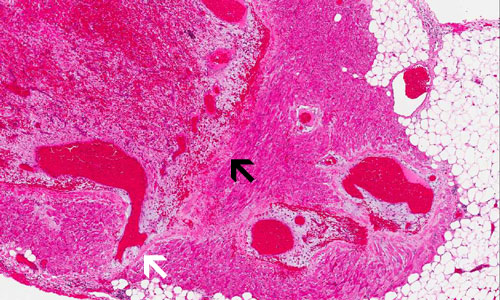 |
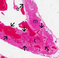
Area 2: Note that in the deepest part of the thrombus (black arrow), the endocardium cannot be well distinguished from either the myocardium or the thrombus. In a location slightly peripheral (white arrow), remnants of the fibrous component of the endocardium can still be recognized. |
 Hematoxylin & eosin |
Area 3: Note that his area probably corresponds to the earliest phase of organizaztion of the throumbus featured by a small amount of fibroblasts and foamy macrophages. |
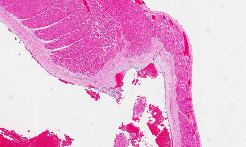
 Hematoxylin & eosin |
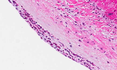
Area 4: A thin layer of polymorphonuclear leukocytes are present in the endocardium. This is most likely acute reactive change due to the procedure but not genuine endocarditis. |
|
History: The specimen was obtained from the cardiac atrium of a 67 year-old woman who has been treated with radiofrequency ablation before for atrial fibrillation (AF).
Histologic Highlights of this Case:
Comment:
|
Original slide is contributed by Dr. Kar-Ming Fung, University of Oklahoma Health Sciences Center, Oklahoma, U.S.A.