History: The patient was a 25 year-old woman who complained of abdominal pain. Laboratory examination demonstrated elevated hCG. The specimen was later obtained.
Histologic Highlights of this Case:
-
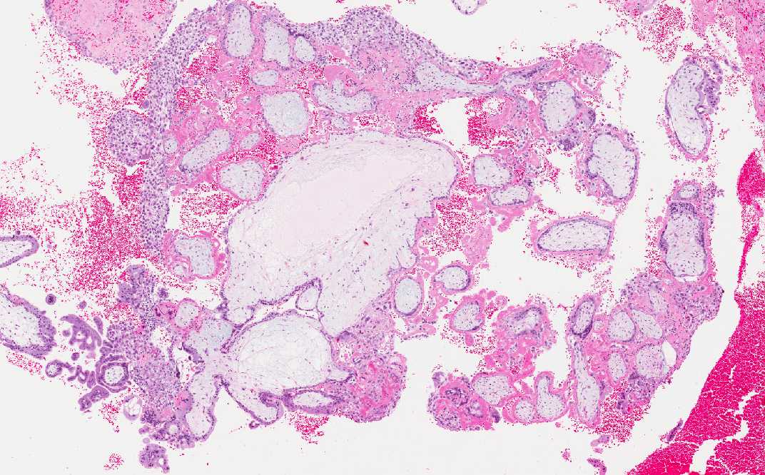
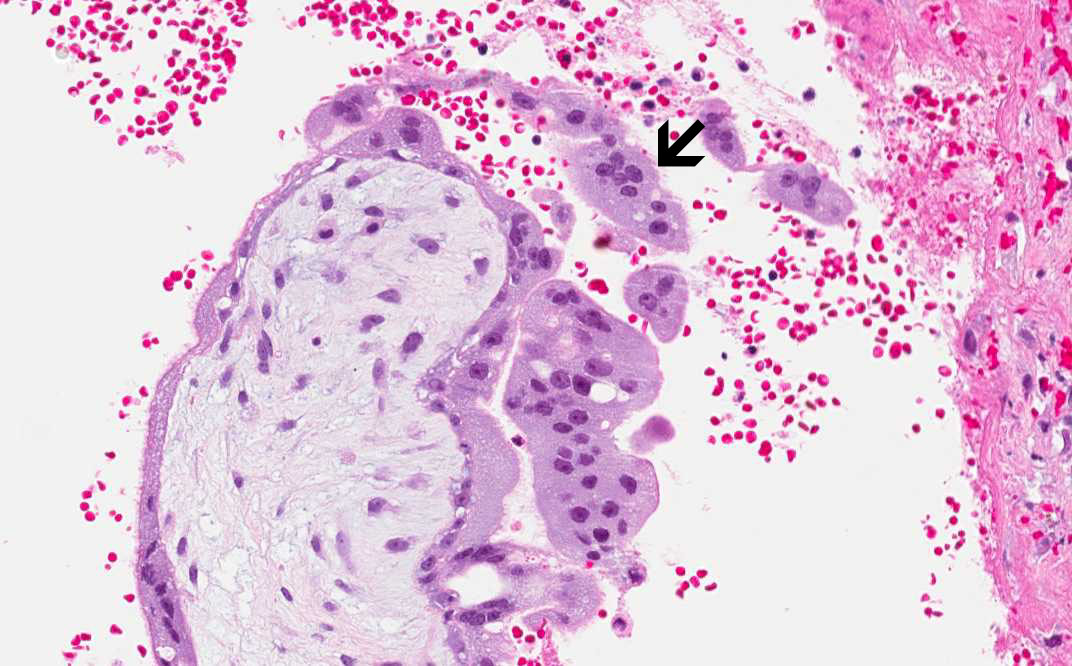
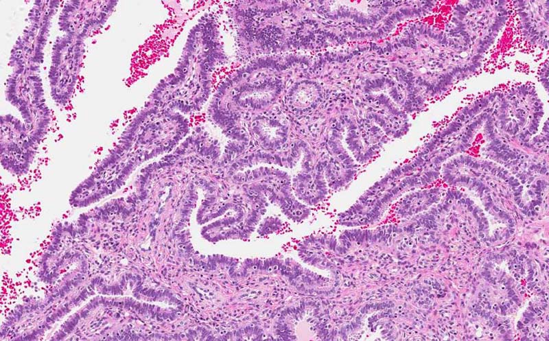
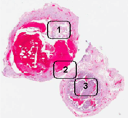
Trophoblastic villi are noted in Area 1 but no fetal tissue is noted. Trophoblastic villi are far more common to be found than fetal tissues in these cases. Note the multinucleated syncytiotrophoblasts (arrow).
-
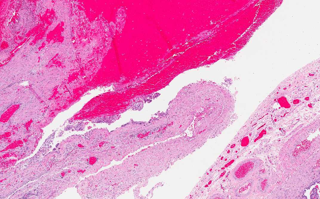
The fallopian tube is filled with fallopian tube and the wall of the fallopian tube is compressed by the hematoma. In some of the cases, the tube may rupture.
-
Note that normal fallopian tube is present in the remaining part of the tube not involved the ectopic pregnancy.