Case No.: G-001
Diagnosis: Prostatic adenocarcinoma (acina, not otherwise specified) with Gleason score 3 and 4 components.
Organ: Prostate
Last Updated: 12/20/2009
|
|
|
Hematoxylin & eosin |
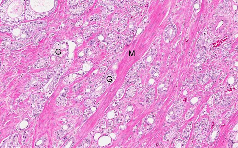
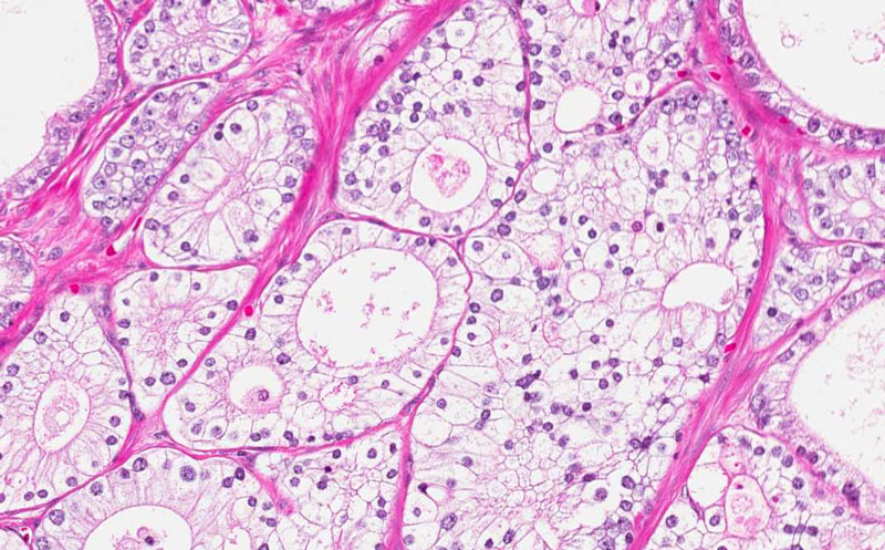
Tumor: The tumor is characterized by small neoplastic acini (G) that infiltrate. In this area, residual smooth muscle fibers (M) are entrapped between the infiltrating neoplastic glands. |
|
Hematoxylin & eosin |
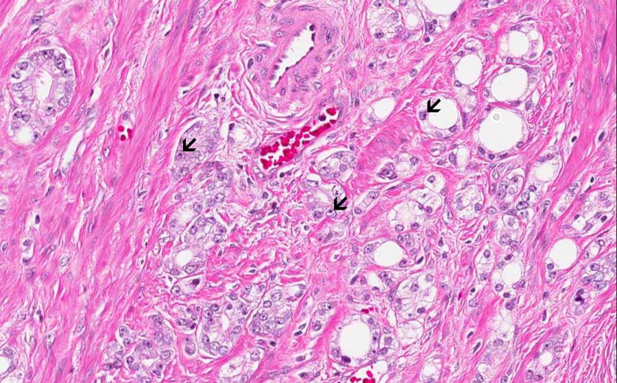
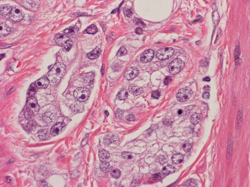
Tumor: The neoplastic glands vary from round with a lumen to some glands that look like fusion of two glands. Note the prominent nucleoli of the carcinoma cells (arrow) and the clear cytoplasm. |
|
Hematoxylin & eosin |
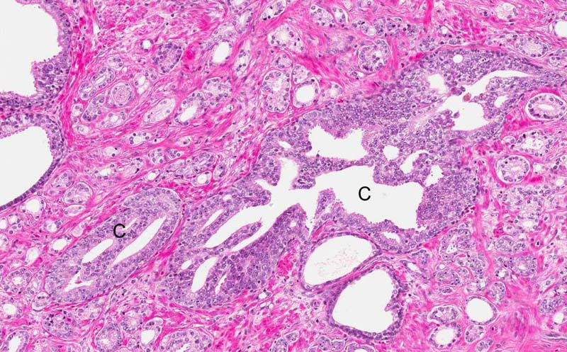
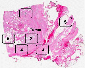
Area 1: In some of the tumor, a cribiform growth pattern is also present (C). |
|
Hematoxylin & eosin |
Area 2: Another area with cribiform area. |
|
Hematoxylin & eosin |
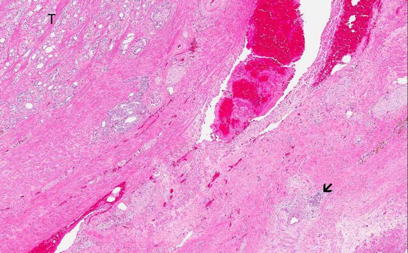
Area 3: This tumor is widely infiltrative. Note the infiltrating focus (arrow) is rather far away from the main tumor (T). |
|
Hematoxylin & eosin |
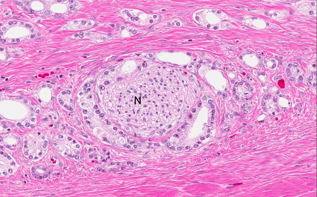
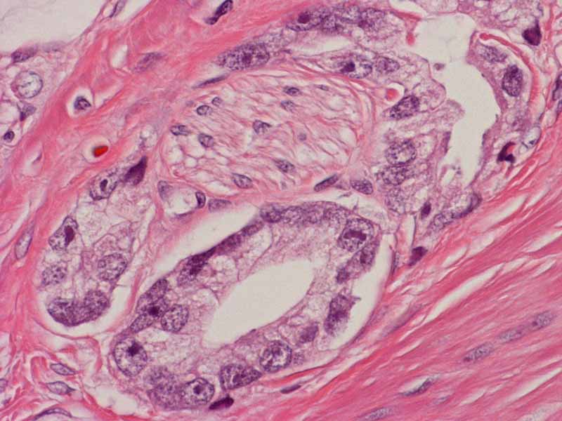
Area 4: Perineural invasion (invasion along the perineural space) is noted in this tumor. Note the peripheral nerve (N). |
|
Hematoxylin & eosin |
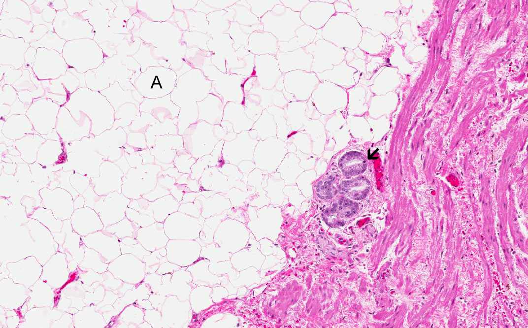
Area 5: This tumor has extracapsular invasion (arrow) into the periprostatic adipose tissue (A). |
|
Hematoxylin & eosin |
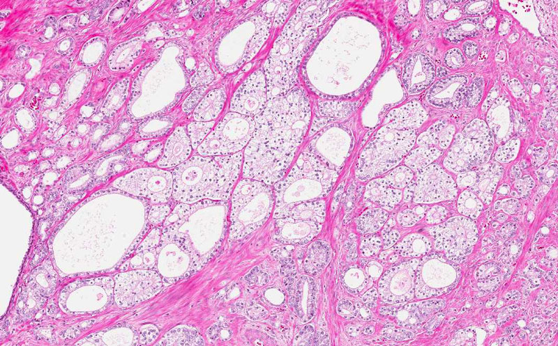
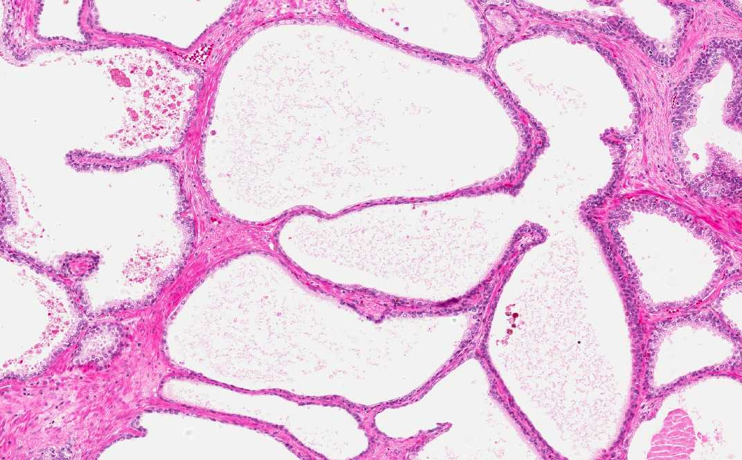
Area 6: This are residual prostatic glands with dilatation and atrophic changes of the epithelium. |
|
History: The patient is a 67 year-old man with increased prostatic specific antigen (PSA). Needle biopsy of the prostate gland reviewed adenocacinoma. A radical radical prostatectomy was performed and yielded the current specimen.
Histologic Highlights of this Case:
|
Bonus images:
Hematoxylin & eosin |
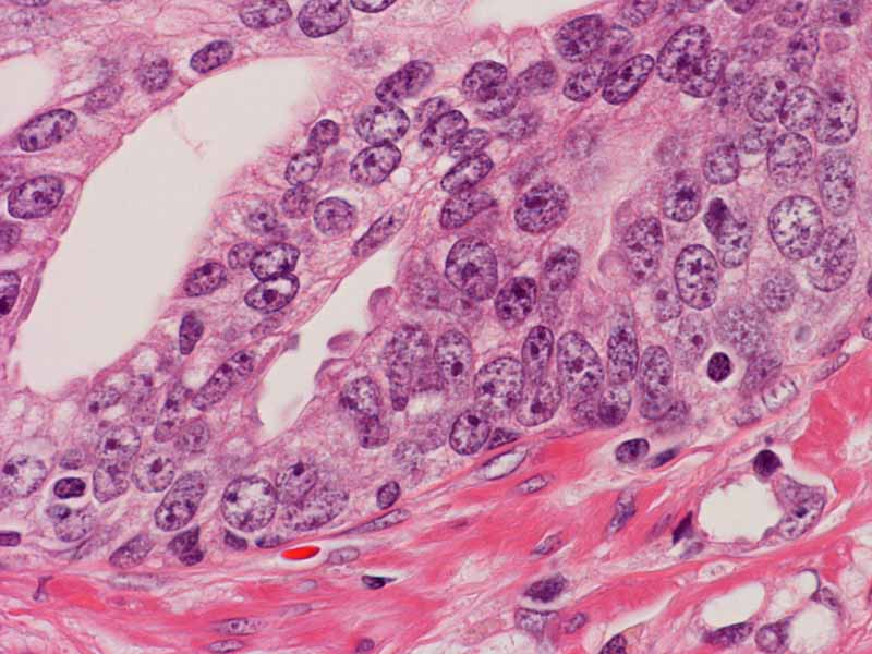
These are additional high-magnification images to show nuclear details. Note the enlarged nuclei. |
Original slide is contributed by Dr. Kar-Ming Fung, University of Oklahoma Health Sciences Center, Oklahoma, U.S.A.