Case No.: H-001
Diagnosis: Sarcoidosis
Organ: Lymph node, neck
Last Updated: 08/21/2010
|
|
|
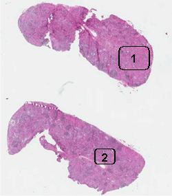
Hematoxylin & eosin |
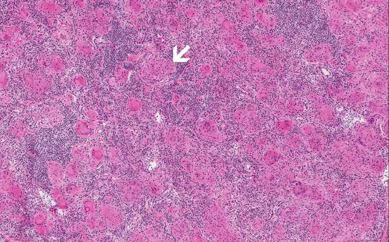
Area 1: Granulomas (arrow) are collections of histiocytes. These cells will be strongly reactive for antibody against CD68 or CD163 which mark histiocytes. Immunohistochemistry is useful when the granuloma is not as well defined or if there is a question that these cells may represent metastatic carcinoma. Granulomas in sarcoidosis is usually free of necrosis. Typically, they are not infiltrated by a substantial amount of lymphocytes and they are quite well defined from the surrounding lymphocytes. They also are more likely to be round or oval. In contrast, granulomas in tuberculosis are usually infiltrated by lymphocytes, much less well defined, more irregular in shape, and may have caseous necrosis. |
|
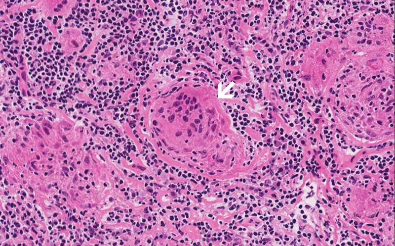
Hematoxylin & eosin |
Area 2: Asteroid bodies (arrow) can be seen in this giant cell. |
|
History: The patient was a 38 year-old African American woman who presented with a painless neck nodule. The nodule was non-tender and the patient could not clearly recall when did she recognized the nodule but it had been there for some time as per the patient. No constitutional symptoms were reported by the patient. The patient has no history of occupational exposure to beryllium or other chemicals. A CT scan was performed which demonstrated multiple enlarged lymph nodes in the mediastinum. The differential diagnoses include lymphoma, metastatic carcinoma, mycobacterial and fungal infection, sarcoidosis and other non-infectious granulomatous disease. The nodule in the neck was excised. The specimen was that of a lymph node, 1.8 cm in greatest dimension, with a solid cut surface. A touch preparation was performed and there was no evidence of lymphoma or metastatic carcinoma. The specimen was submitted for histologic examination. The angiotensin-converting enzyme (ACE) level of this patient was elevated.
Histologic Highlights of this Case:
Comment: Sarcoidosis, also known as Besnier-Boeck disease is a disease characterized by the formation of granulomas in multiple organs particularly the lung and lymph nodes. Typically, these granulomas are non-necrotizing. Many of these lesions are asymptomatic and are discovered incidentally which would then raise a question of a neoplasm particularly when a solitary lesion is identified. Since graunulomas arising from multiple organs is a non-specific findings and can be seen in multiple conditions, sarcoidosis is a clinical pathological diagnosis which requires correlation of both clinical symptoms, laboratory findings, and pathologic examination. Clinical imaging is very helpful. It is also important to check out the occupational history. Angiotensin-converting enzyme (ACE) is usually elevated in sarcoidosis. Pathologic examination and micorbiological culture can help to rule out fungal or acid fast bacilli infection as well as other mimickers such as lymphoma, lymphadenopathy, metastatic tumor, Wegener's granulomatosis and others. |
Original slide is contributed by Dr. Kar-Ming Fung, University of Oklahoma Health Sciences Center, Oklahoma, U.S.A.