History: The patient was a 57 year-old woman presented for evaluation of an left groin mass. The mass was asymptomatic and painless. She reported a weight loss of 30 pounds in 5 months. On physical examination, there were also enlarged axillary lymph node. A biopsy of the inguinal mass was performed and yielded the current specimen.
Histologic Highlights of this Case:
-
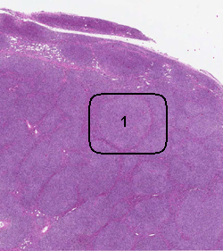
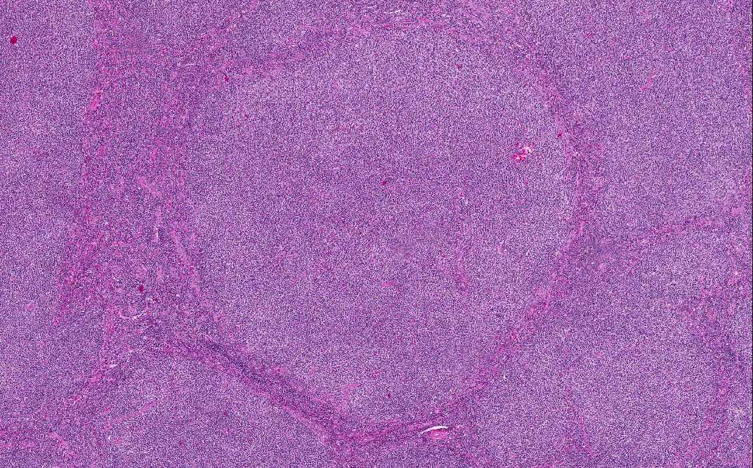
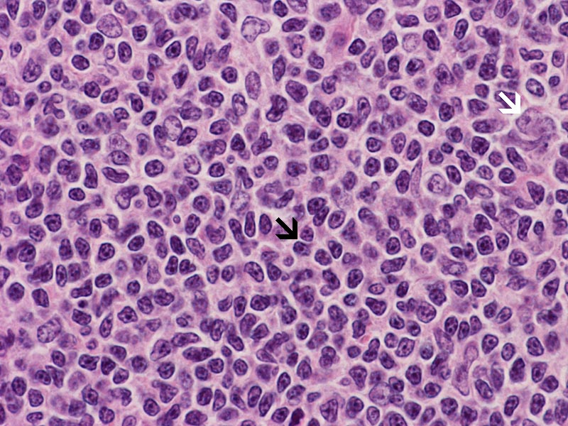
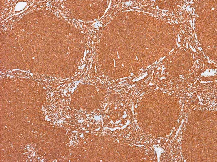
On scanning magnification, the lymph node demonstrates total effacement of the architecture. It is replaced by neoplastic follicles of uniform size, showing closely packed pattern. The follicles lack mantle zone as would be expected in normal lymph node. This is a classic pattern of follicular lymphoma. The follicles are composed of densely packed centrocytes and centroblastes without a “starry-sky” pattern or polarization. In particular, specimen, no no areas with diffuse arrangement is noted. The thin rim of lymphocytes around the follicles are mostly non-neoplastic T cells.
-
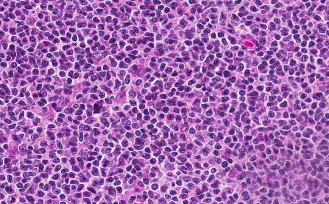
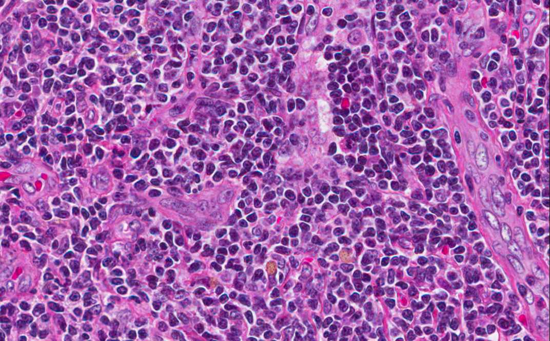
Cytologically, the neoplastic lymphocytes (centrocytes) are small to medium size with angulated, elongated, cleaved, or twisted nuclei. Chromatin is evenly dispersed, small nucleoli may be seen. Centrocytes appear more monomorphic than lymphocytes of the normal lymphoid follicles. The nuclear pleomorphism is low. Rare centroblasts are seen. Centroblasts are larger cells containing vesicular nuclei with one to three basophilic nuclei apposing nuclear membrane. These are the features of low-grade follicular lymphoma.
Immunohistochemistry:
-
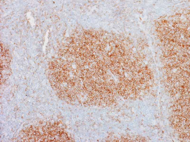
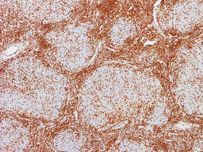
The tumor cells are positive for CD20, CD10 and Bcl-2 but negative for CD3 and other T-cell markers (see below).
Additional Information:
-
Flow cytometry was performed and showed B-cell population with kappa restriction consistent with B-cell lymphoma.
-
The degree of follicular formation can varies from predominantly follicular (<75%) to minimally follicullar (<25%).
-
Histologic grade can vary from low to high. In the WHO system, there are 3 histologic grade with grade 3 the highest. The histologic grade is determined by the proportion of centroblasts.
-
Over 80% of follicular lymphoma have t(14;18) translocation. Bcl-2 protein can be demonstrated in neoplastic germinal centers by immunohistochemical stain in majority of grade 1-2 follicular lymphoma (85-97%). Bcl-2 protein is expressed by B and T lymphocytes but it is never normal to see BCL2 expression in benign germinal center.
-
Molecular detection of immunoglobin heavy chain rearrangment (IgH) is a good help to confirm monoclonality in lymphoma.