Case No.: K-003 Quiz
Diagnosis: Adrenocortical adenoma
Organ: Adrenal gland
Last Updated: 3/21/2011
|
|
|
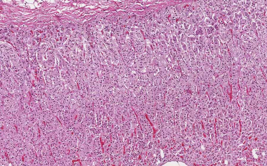
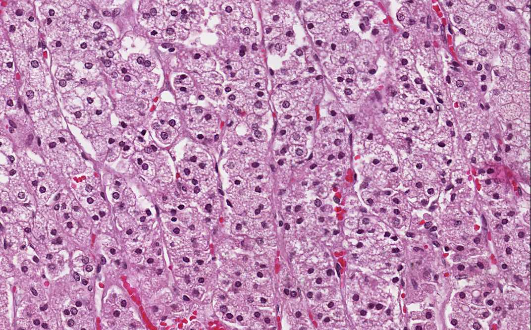
Hematoxylin & eosin |
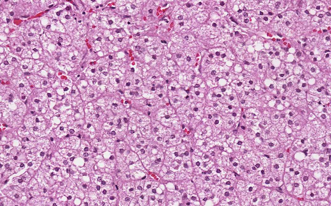
Area 1: Starting from the capsule, the adrenal cortex is composed of three layers or zones, namely the zona glomerulosa, zona fasciculata, and zona reticularis. The zona fasciculata is the thickest and comprises the bulk of what is being shown in this image. The cells are polygonal and monotonous. The nuclei are small, round, centrally located and the cytoplasm is foamy. Compare these cells with the tumor cells from Area 3. |
|
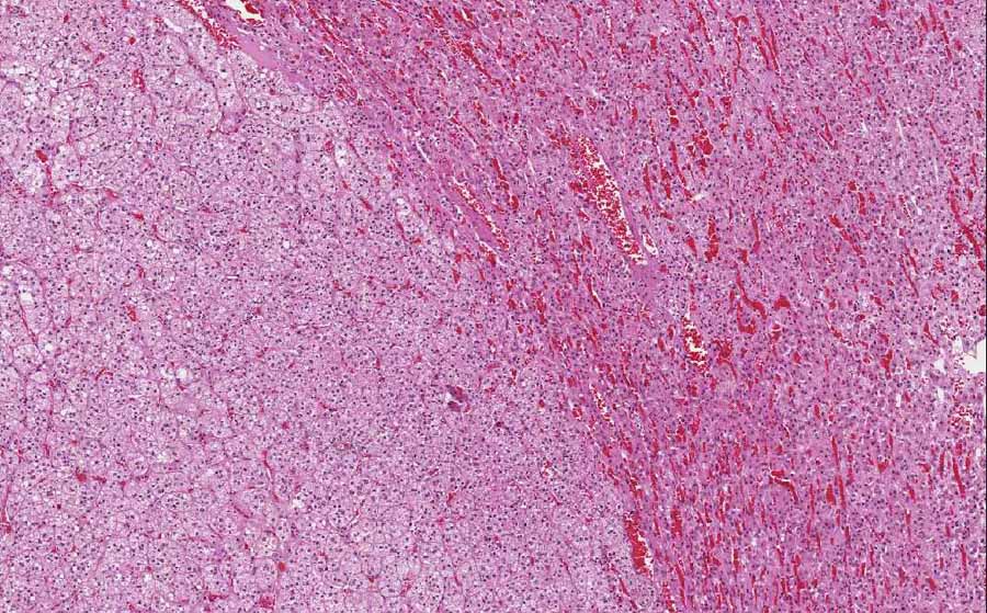
Hematoxylin & eosin |
Area 2: This is the interface between the tumor and the residual adrenal cells. Note that the residual cells around the tumor have more eosinophilic cytoplasm and are less foamy. |
|
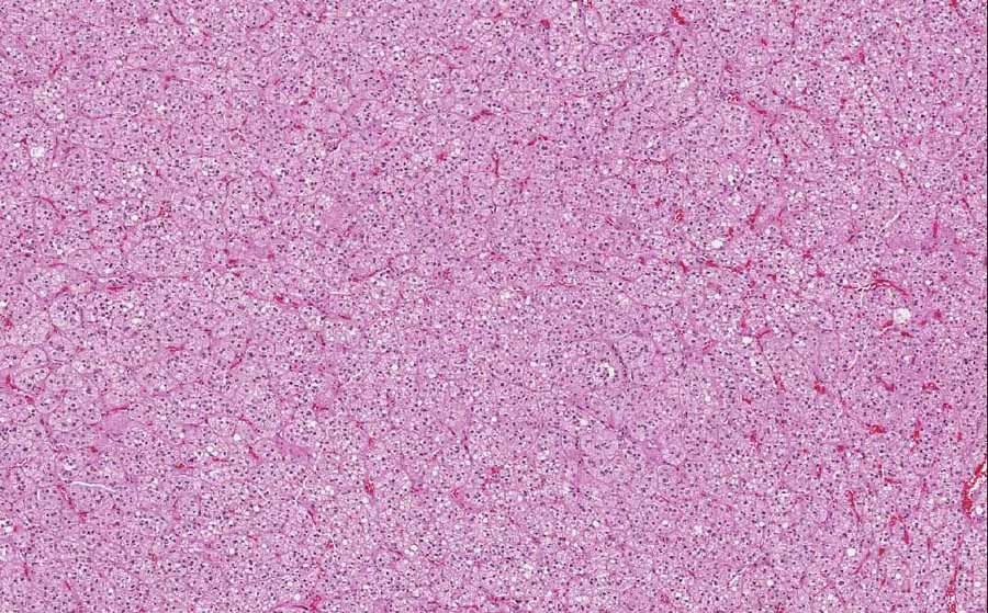
Hematoxylin & eosin |
Area 3: The tumor has close resemblance to the residual adrenal cortex in terms of both histologic architecture and cytoplasmic features. |
|
History: The patient was a 62 year-old man who presented to the hospital with a history of left flank pain of unknown duration and recent hematuria. An MRI studied demonstrated a 3.4 cm solid mass in his left kidney. A radical nephrectomy was performed and the mass was proved to be a clear cell renal cell carcinoma. The adrenal gland was also removed and a small 2 cm solid nodule was noted. The image being shown here was obtained from the adrenal nodule.
Gross Pathology: The tumor is 2.2 cm in greatest dimenstion. It is in the form of a single, well-demarcated nodule surrounded by a thin layer of normal appearing adrenal cortex. The tumor has a pale yellow, solid cut surface without any hemorrhage or necrosis. The residual adrenal cortex does not show any evidence of atrophy.
Histologic Highlights of this Case:
Comment: Adrenocortical adenomas are often, but not always, incidental finding particularly when they are small and non-functional. These tumor can be functional or non-functional and morphologic features alone cannot determine whether they are functional. Clinical assessment and laboratory measurement of the hormone level is necessary. However, functional adrenocortical adenomas are often associated with atrophy in the associated adrenal cortex and in the contralateral adrenal gland. |
Original slide is contributed by Pathology Learning Center, University of Iowa (Iowa Image Collection)