Online Slide/Full Screen
Open with ImageScope
|
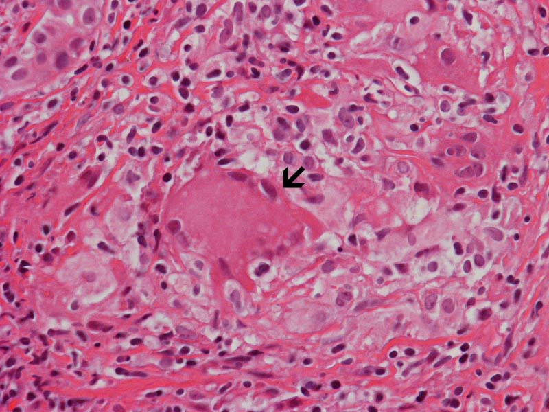
Hematoxylin & eosin |
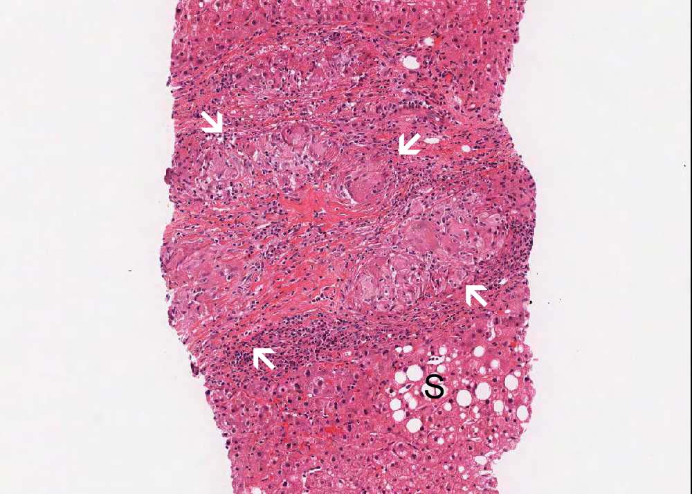
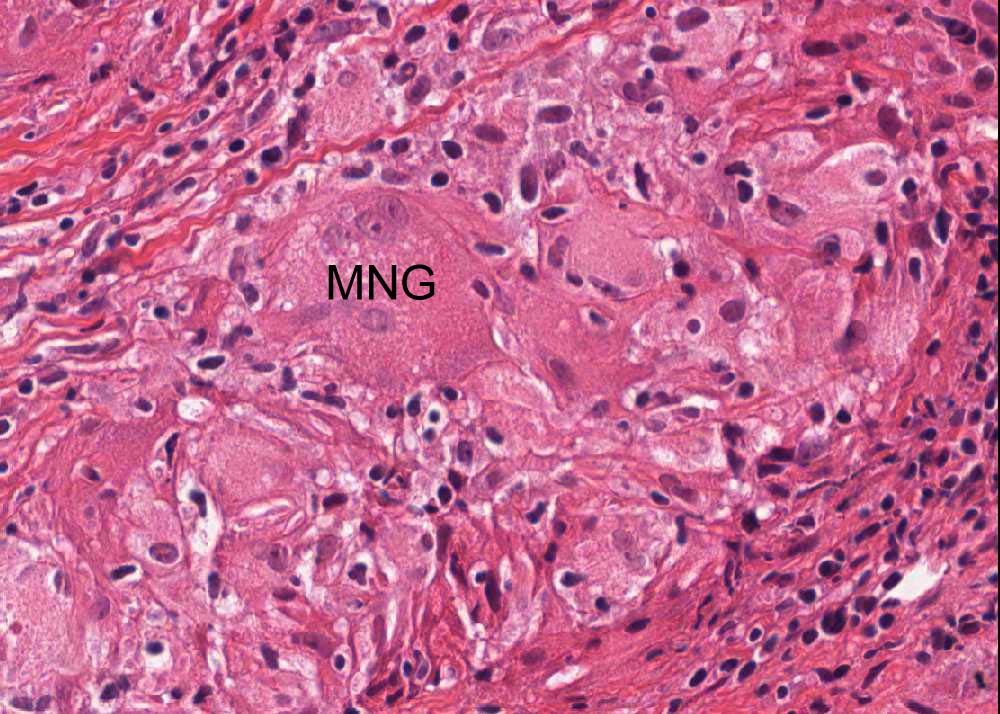
Area 1: The granuloma is outlined by the arrows and the steatosis (S) is also present. Multinucleated giant cells (MNG) is present. Note that there is no necrosis in these granulomas. Note that the granuloma is surrounded by a small amount of reactive lymphocytes. |
|
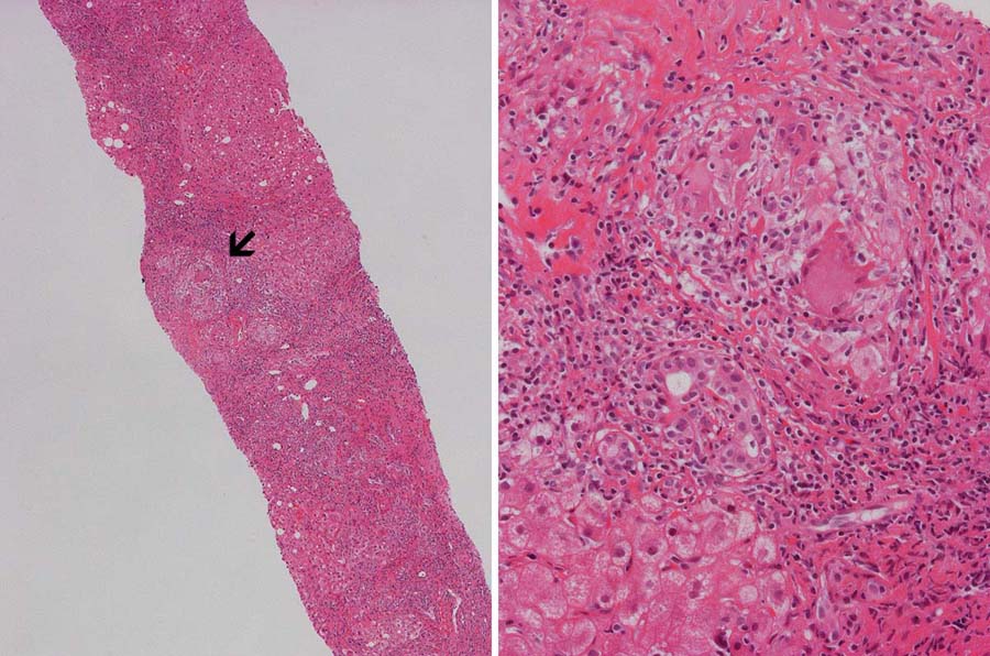
Hematoxylin & eosin |
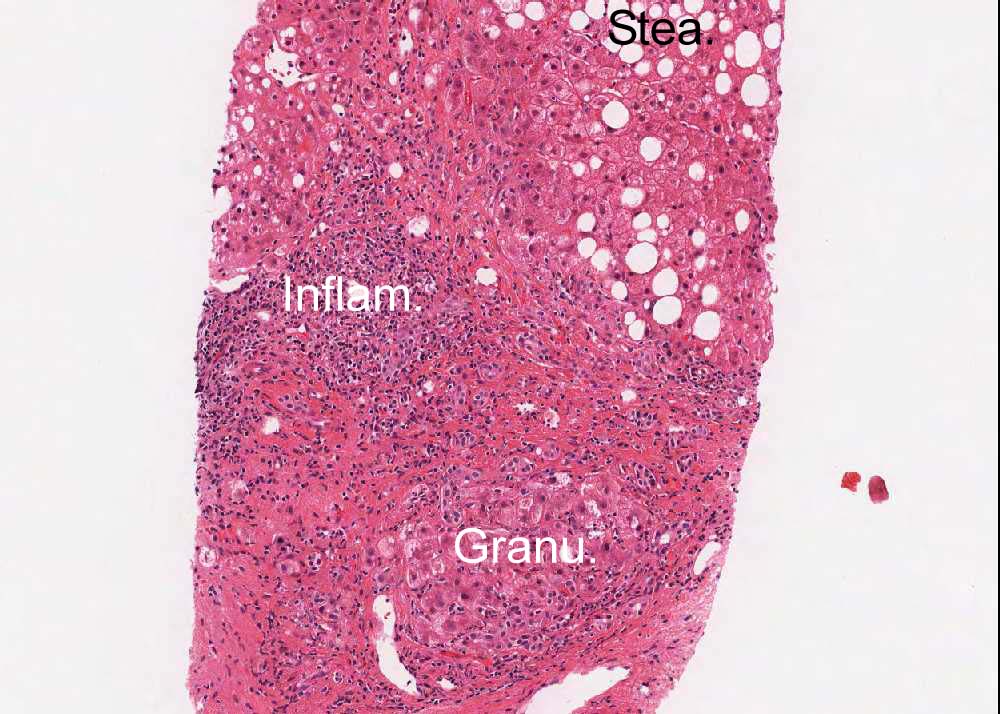
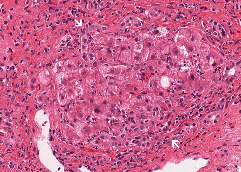
Area 2: The granuloma (Granu.) is surrounded by lymphocytes. Note that there are also areas with mainly lymphocytic infiltration and the granulomatous changes is subtle (Inflam.). Steatosis (Stea.) is also present. Note that the non-steatotic hepatocytes in the steatotic area has more regular cell shape in comparison to the histiocytes. In some areas as illustrated here in the high-magnification, residual hepatocytes may be difficult to be distinguished from granuloma base on morphology alone (arrows). |
|
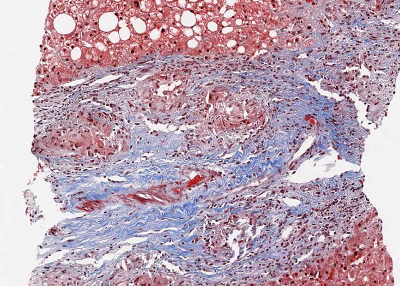
Trichrome |
Area 3: The granuloma is surround by fibrous tissue which is well demonstrated by the Masson's trichrome stain which stains collagenous fibrous area blue. Click on the online slide to compare with the hematoxylin and eosin stained slide. |