Case No.: N-013
Diagnosis: Gangliocytic paraganglioma
Organ: Spinal cord, filum terminale
Last Updated: 07/05/2010
|
|
|
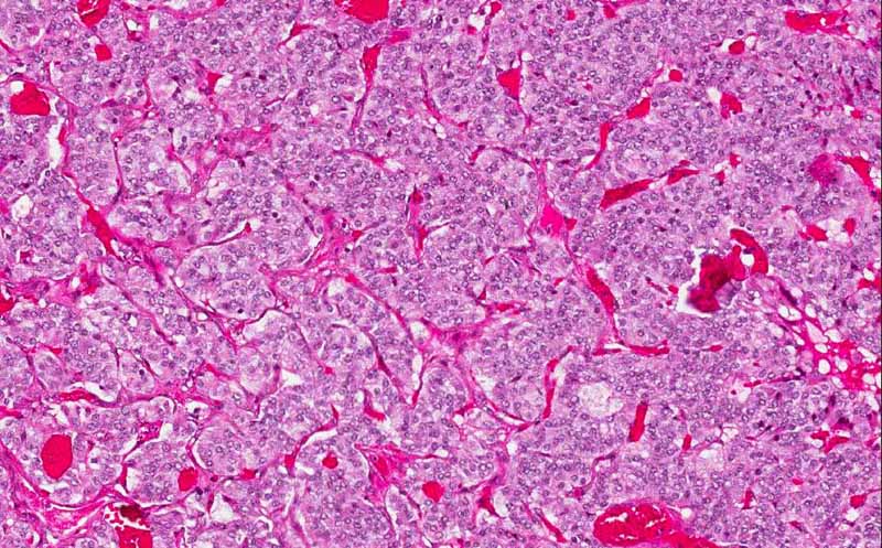
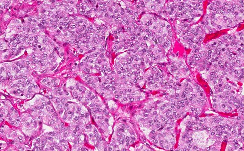
Hematoxylin & eosin |
Area 1: Medium and high magnification of the organoid "Zellballen" cell nests. Note that the nest are surrounded by a network of fine capillaries. |
|
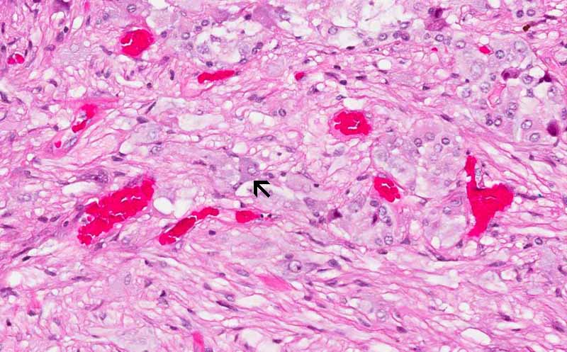
Hematoxylin & eosin |
Area 2: Ganglionic neurons are present (arrow) in some areas of this tumor. Note that the ganglionic cells are embeded in a schwannian background. |
|
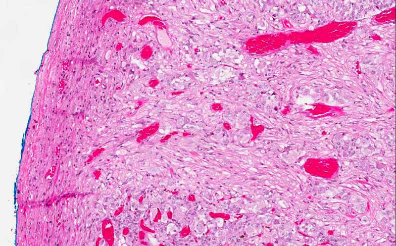
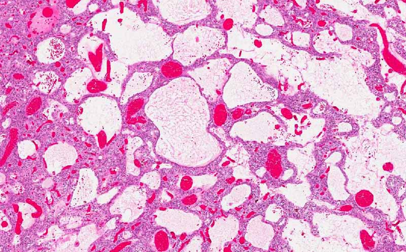
Hematoxylin & eosin |
Area 3: Dilated structures most consistent cystic changes or dilated blood vessels. |
|
History: The patient was a 40 year-old man who complained of chronic back pain and is getting severe in the lower lumbar spine and sacrum with radiation to the right lower extremity and left lower extremity. The quality is constant ache with intermittent shooting pain. The pain got worse on lying down and is not relieved by anything. MRI scan demonstrated a 2 cm well defined, oval spinal mass in the distal end of the spinal cord. The mass was excised and yielded the current specimen.
Capsule Summary: Paraganglioma is usually a benign tumor but malignant ones may occur. It usually occur in area where paraganglia are found and these tumors displays features of paraganglia. One of the two salient features is the characteristic organoid “Zellballen” or cell nests that mimic chief cells. The other characteristic is that these tumor is composed predominantly of a population of large cells that are positive for neuroendocrine markers such as synaptophysin or chromogranin. These cells bear features of the chief cells. There is also a much smaller population of sustentacular cells which are negative for neuroendocrine markers but positive for S100. When gangliocytic cells is part of the tumor, these tumors are termed gangliocytic paraganglioma as in this case.
Histologic Highlights of this Case:
|
Bonus Images:
|
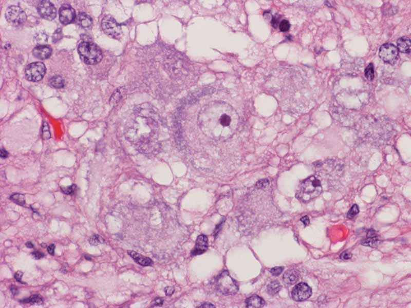
Hematoxylin & eosin |
Ganglionic cells: This is a high magnification photo taken from the ganglionic cells of this cases. Note the large size of the cell, the abundant amount of cytoplasm, the polygonal to oval shape, the bluish rim of staining in the cytoplasm, the large nuclei and the prominent nucleoli. All are features of a ganglionic cells. |
|
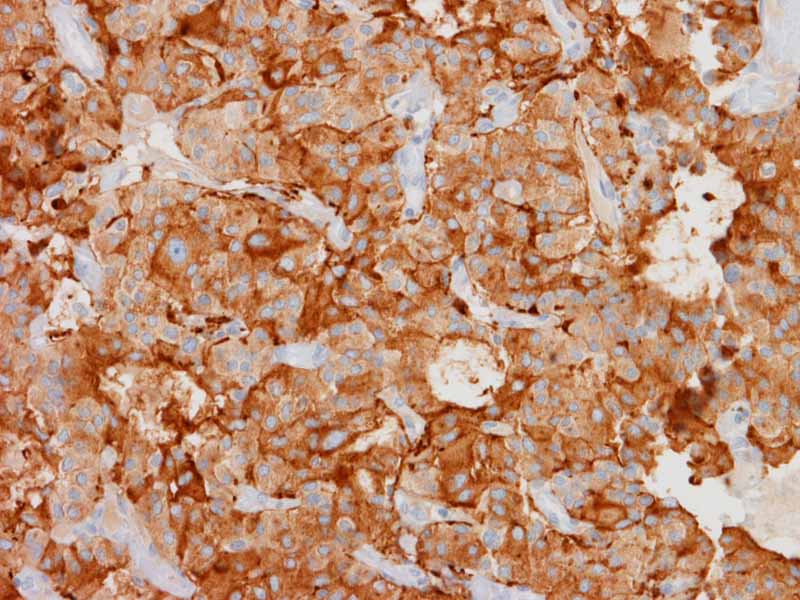
Synaptophysin |
Synaptophysin positive cells: Immunohistochemistry demonstrate strong immunoreactivity for synatophysin in the large tumor cells. Synaptophysin is a marker for neuroendocrine differentiation. These tumor cells have morphological and immunohistochemical features of normal chief cells in paraganglia. |
|
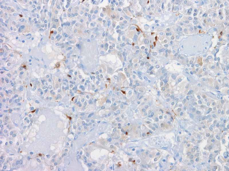
S100 |
Sustentacular cells: The sustentacular cells are positive for S100 which allows rapid identification. Note that the sustentacular cells are small compared to the tumor cells in the cell nests. |
Original slide is contributed by Dr. Kar-Ming Fung, University of Oklahoma Health Sciences Center, Oklahoma, U.S.A.