Case No.: N-024
Diagnosis: Abscess
Organ: Brain
Last Updated: 3/21/2011
|
|
|
Hematoxylin & eosin |
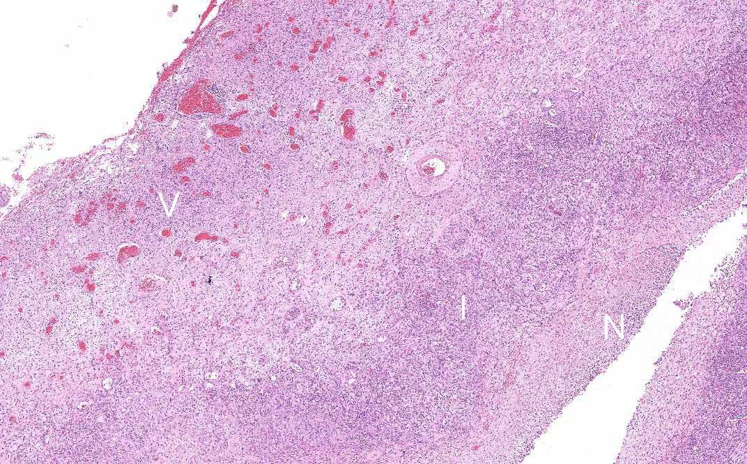
Area 1: The wall of the abscess is composed of several layers. The inner most layer is coated by the necrotic debris (N). There is intense acute inflammatory cell infiltration in the layer immediately under the necrotic debris (I). There is prominent vascular proliferation in the deeper layer (V). The increase in cellularity is partially due to gliiosis as well as inflammatory cell infiltration. The inflammatory cells are composed predominantly of lymphocytes and some eosinophils. There are not that much neutrophils. One of the most likely possibility is that the patient has been treated with antibiotics and the acute inflammation has already subsided. Many foamy macrophages are also present. |
|
Hematoxylin & eosin |
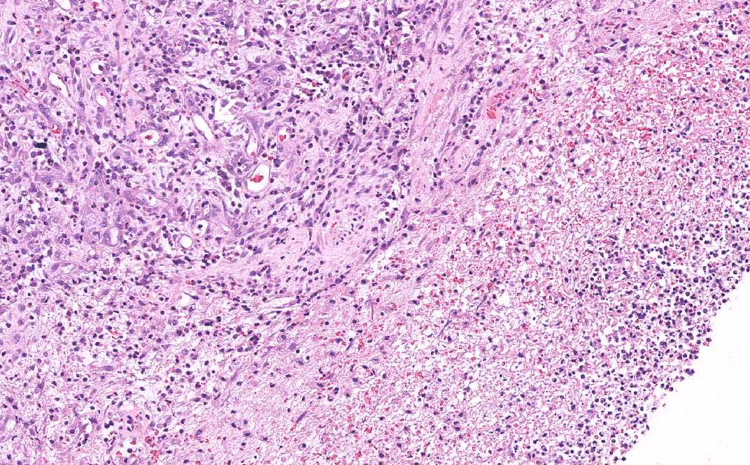
Area 1: In the layer with intense inflammatory cell infiltration, there is also substantial vascular proliferation. These are neovascularizations and they do not have large lumens. |
|
Hematoxylin & eosin |
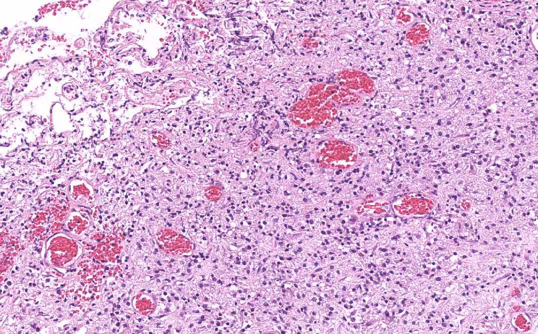
Area 1: In contrast to the outer layer of the abscess, the blood vessels are more mature and have dilated lumens. |
|
History: This slide is taken from the archive and the history was not known. This condition typically appears as an enhancing cystic lesion on MRI (ring enhancement). It can occur in all age group and has increased incidence in patient with congenital heart disease particularly those with right to left shunt.
Histologic Highlights of this Case:
|
Original slide is contributed by Fred R Dee MD, Department of Pathology, University of Iowa. (Iowa Image Collection)