Case No.: N-023
Diagnosis: X-linked adrenoleukodystrophy
Organ: Cerebellum
Last Updated: 3/21/2011
|
|
|
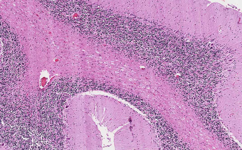
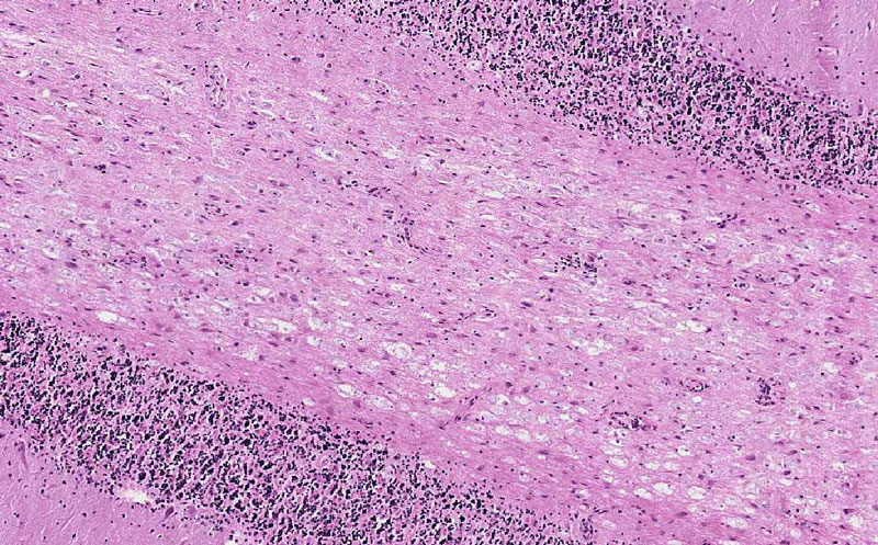
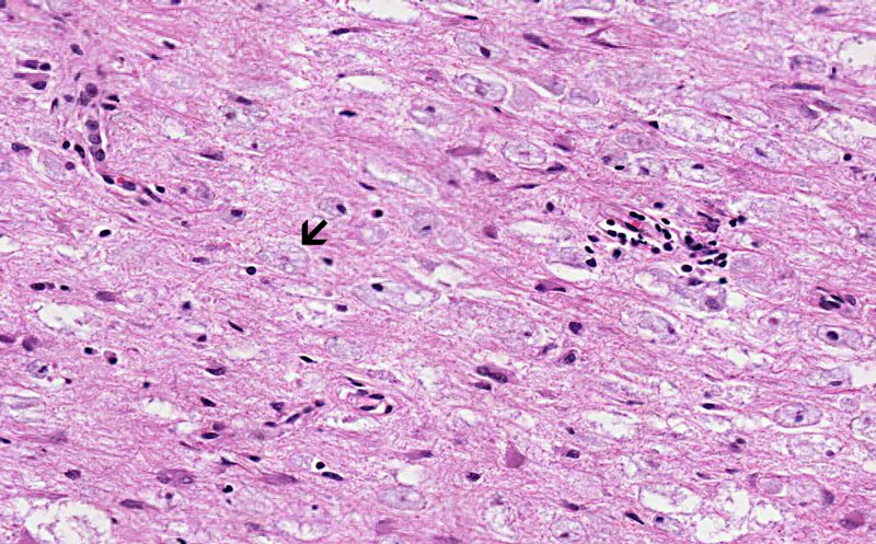
Hematoxylin & eosin |
Area 1: There are many histiocytes with grayish, sometime foamy cytoplasm. |
|
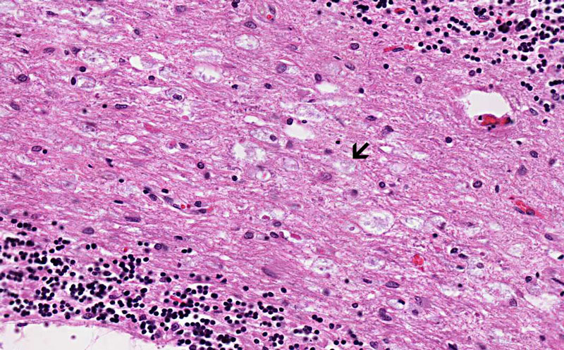
Hematoxylin & eosin |
Area 2: The density of infiltrating histiocytes is much higher here. Note that there is no chronic inflammatory cell infiltration. |
|
History: This slide was taken from the archive and the history was not provided. This specimen was obtained from the autopsy of a case with know x-linked adrenoleukodystrophy.
Histologic Highlights of this Case:
Comment:
|
Bonus Images:
|
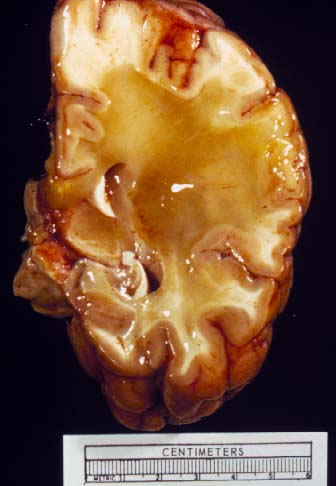
Gross Photo |
Gross Photo: This gross photo was taken from a different case. Note that the white matter is diffusely replaced by a yellow gelatin like substance. |
|
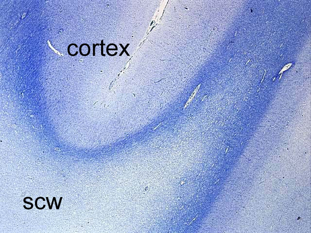
Luxol Bast Blue-Cresyl Violet |
Loss of myelin: Myelin is staind blue in Luxol fast blue-Cresyl violet preparation. This image is taken from the cerebral cortex and subcortical white matter from a different case of x-linked adrenoleukodystrophy. Note that there is extensive loss of myelin in the deeper part of the subcortical white matter (scw). Myelin in the subcortical fibers are preserved. |
|
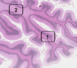
Hematoxylin-Eosin |
 Luxol fast blue-Cresyl violet |
For comparison: These virtual slides are obtained from the cerebellum of an adult without neurologic pathologic changes. You can compare the pattern of myelination with the image above. |
Original slide is contributed by Pathology Learning Center, University of Iowa (Iowa Image Collection).