Case No.: S-001

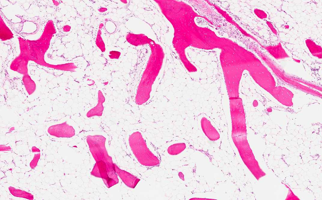
Diagnosis: Osteolipoma (lipoma with ossification)

Organ: Soft tissue, Ankle

Last Updated: 08/21/2010
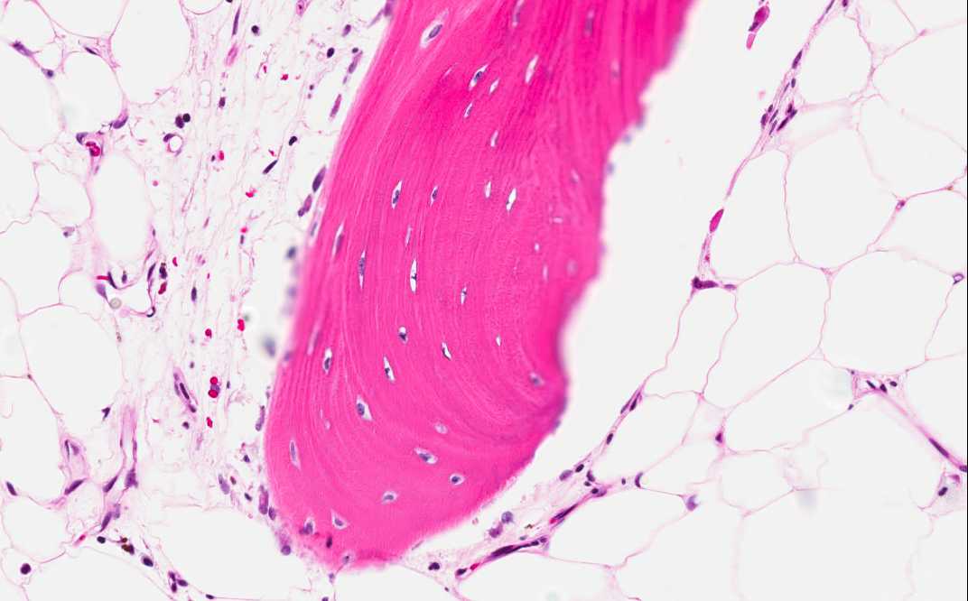
 

Online Slide/Full Screen/Open with ImageScope

Hematoxylin & eosin

Area 1: Note that the bone at the internal part of the nodule is composed of mature, lamellar bone.
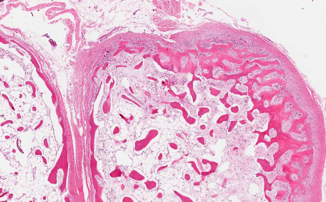
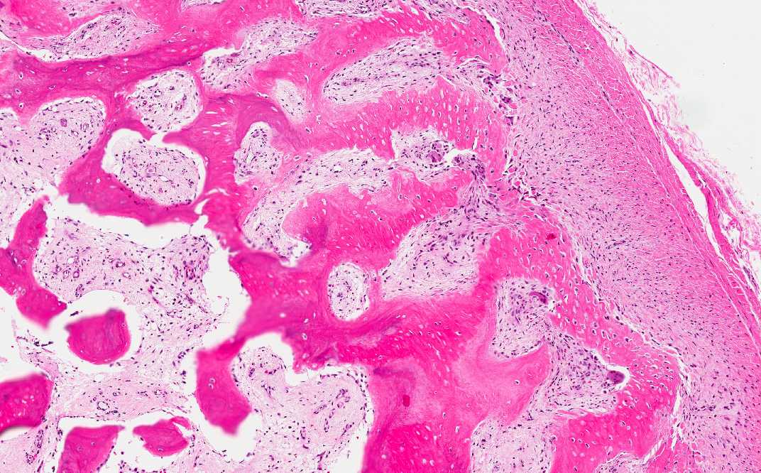
Hematoxylin & eosin

Area 2: These images illustrate the ossifying process of the shell of the lesion.

History:  The patient was a 44 year-old woman who complained of a 3 cm hard mass in her ankle at the medial aspect of her tibia. She had a mass that was removed from that area approximately about 3 years ago and the diagnosis was lipoma. She developed postoperative infection with a prolonged course and was treated. On physical examination, the mass was hard like a stone and was thought to be mineralized. MRI revealed fat density in the mass. The current mass was excised and yielded the this specimen.


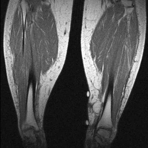
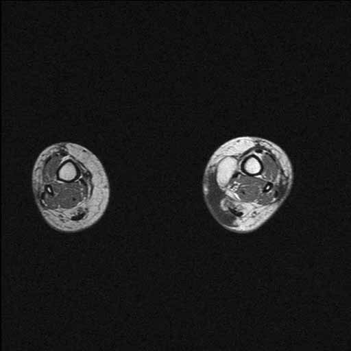
MRI: There is an oval mass with fat density in the left ankle. The mass is very well demarcated from the surrounding tissue without evidence of invasion into the surrounding tissue.

MRI


Gross Pathology:

  • The specimen has the shape of a round to oval smooth, bony hard nodule of 2.9 cm in greatest dimension. The entire surface is covered by a thin layer of thin fibrous tissue. There is no exudate or hemorrhage on the surface. The nodule can be easily cut through with a saw. The cut surface resemble bone but is less dense. Sections are decalcified and submitted for histologic examination.

Histologic Highlights of this Case: 

  • The nodule is composed of a calcified shell of bone and scant islands of cartilage (not shown in this image). The internal part of the nodule is composed exclusively of mature adipose tissue and calcifying bone. No fibrous proliferation of inflammatory cells are present. No mature bone marrow elements are found.

Comment: 

  • Although the overall pathologic findings are suggestive of myositis ossificans, there is no fibrous tissue proliferation at the internal part of the nodule. Instead, there is only mature adipose tissue.

  • The overall finding is most consistent with a lipoma with ossification, also known as osteolipoma. Although lipoma is a fairly common condition, osteolipoma is quite uncommon. They are more likely to occur if there is local irritation such as inflammation around the lipoma.

Original slide is contributed by Dr. Kar-Ming Fung, University of Oklahoma Health Sciences Center, Oklahoma, U.S.A.

 Home Page