|
History: The patient was a 25
year-old woman with a 6 cm mass in her T8 spinal nerve root. The mass
was excised and yielded the specimen being shown here.
Histologic Highlights of this Case:
-
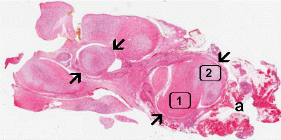
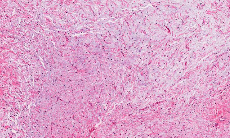
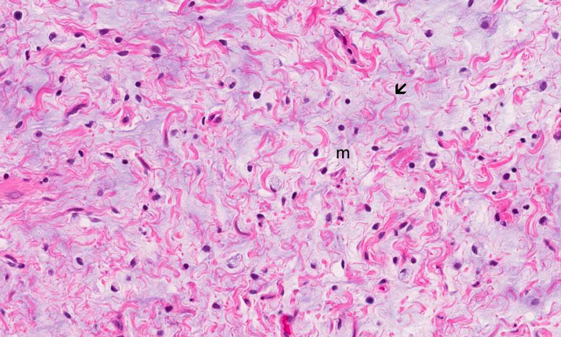
The salient feature of this slide is
that of a spindle cell proliferation with nodule formation (arrows).
The histopathologic picture is reminiscent of the cross section of a
bunch of spaghetti. Some of the area looks more eosinophilic on
scanning magnification (Area 1) and they corresponds to the area
with minimal or no myxomatous changes. These areas contain
substantial amount of collagen and a low density of tumor cells. The
pale areas (Area 2) is characterized by more impressive myxomatous
changes. Note that the two areas intermingle with other
imperceptibly.
-
Note that tumor has pushing margin and
there is no invasion into the surrounding adipose tissue (a)
-
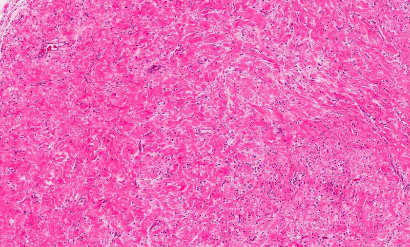
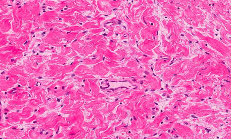
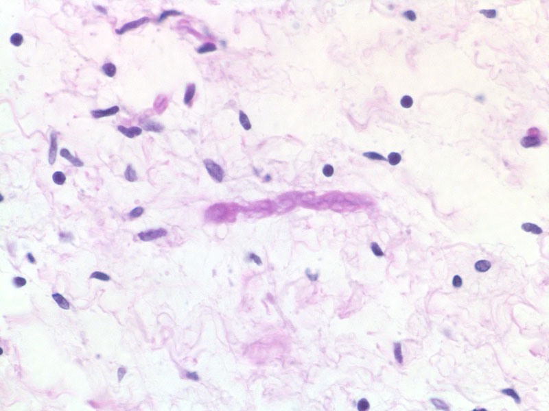
On higher magnification, the tumor is
composed of spindle cells with low small and bland nuclei. There is
no areas with high grade nuclei or active mitosis.
Comment:
-
Neurofibroma tends to occur in the
peripheral part of the peripheral nervous system. A neurofibroma
occurring in the spinal nerve root raises a strong concern for
neurofibromatosis 1 (NF1). Plexiform neurofibroma is seen almost
exclusively in patient with NF1. This patient has a proved history
of NF1 at the time of surgery.
-
Neurofibroma arising in NF1 patient
tends to arise in early age, in usual locations, and has a high
frequency of malignant transformation. A thorough examination of the
specimen and careful search for high grade area is important to rule
the possibility of malignant peripheral nerve sheath tumor.
-
It is important to distinguish
schwannoma from neurofibroma. Neurofibroma characteristically has
myxomatous stroma with spindle cells. While schwannoma tends to push
the nerve to the side, neurofibroma diffusely infiltrates the
affected nerve and therefore entrapped axons can be seen. While they
can be picked up on hematoxylin and eosin stained sections, they are
efficiently detected by using immunohistochemistry that detects the
neurofilament phosphoisoforms that are seen in axons. Silver stains
for axons can also be used. Schwannom tends to be extensively
positive for S100 but neurofibroma are usually only focally
positive.
Unlike schwannoma, neurofibroma usually involve many or all
fascicles of a nerve and make resection impossible. Plexiform
neurofibromas can be found anywhere along the length of the
peripheral nerve. Although the plexiform neurofibroma tends to
confine the bulk of their involvement in large nerves to the nerve
trunks, it also shows diffuse involvement along the length of the
nerve which makes complete excision rather difficult. In contrast,
sporadic neurofibromas are sharply circumscribed.
|