Case No.: T-001
Diagnosis: Papillary carcinoma.
Organ: Thyroid
Last Updated: 12/21/2009
|
|
|
Hematoxylin & eosin |
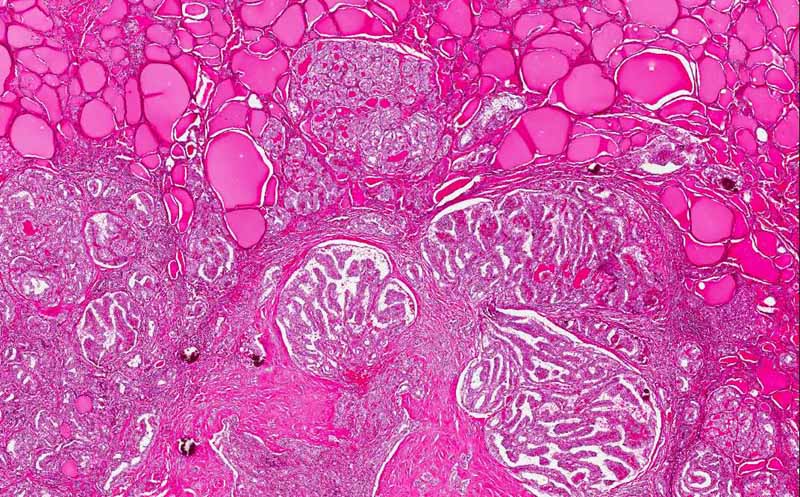
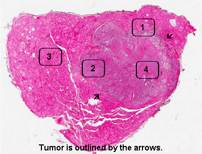
Area 1: Note the irregular margin of the tumor. |
|
Hematoxylin & eosin |
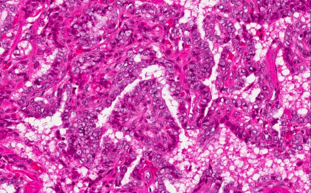
Area 2: Note the papillary growth pattern. It is featured by fine villous or finger like fibrovascular stroma lined by neoplastic epithelial cells. The nuclei are enlarged, often have nuclear grooves and pseudonuclear inclusion. Overlapping of nuclei is also common. |
|
Hematoxylin & eosin |
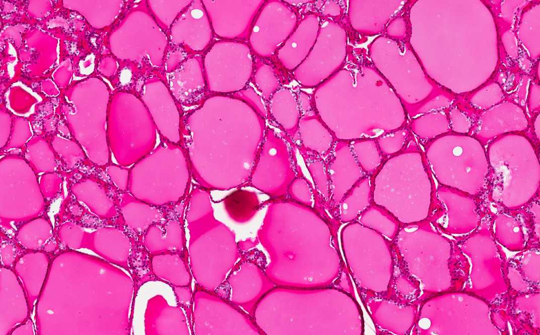
Area 3: This is a normal thyroid area for comparison. |
|
Hematoxylin & eosin |
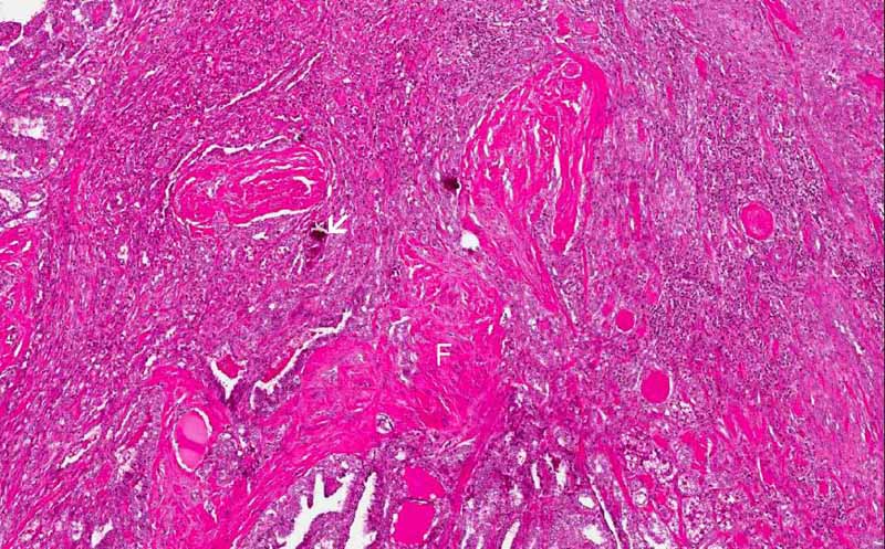
Area 4: Fibrosis (F) and calcifications (C) are characteristic features of thyroid papillary carcinoma. These calcifications can exists as amorphous depositioins or round, concentric psammoma bodies. |
|
History: The patient was a 35 year-old woman who has a firm nodule palpable on her thyroid. A fine need aspiration was performed and reviewed papillary carcinoma of the thyroid. The current slides was obtained from the thyroidectomy specimen.
Histologic Highlights of this Case:
|
Bonus images:
|
Hematoxylin & eosin |
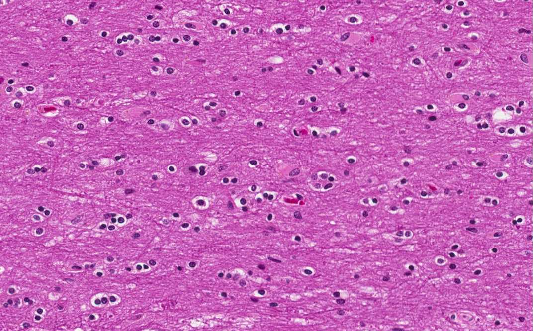
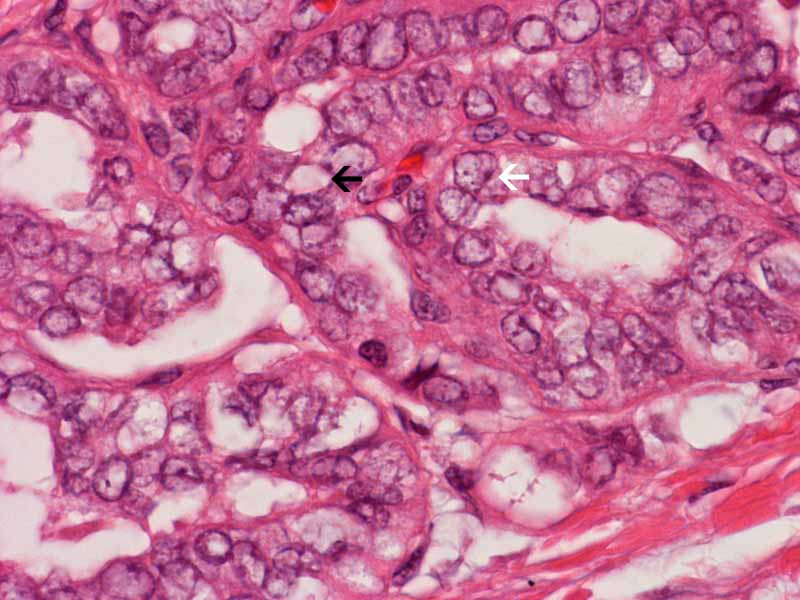
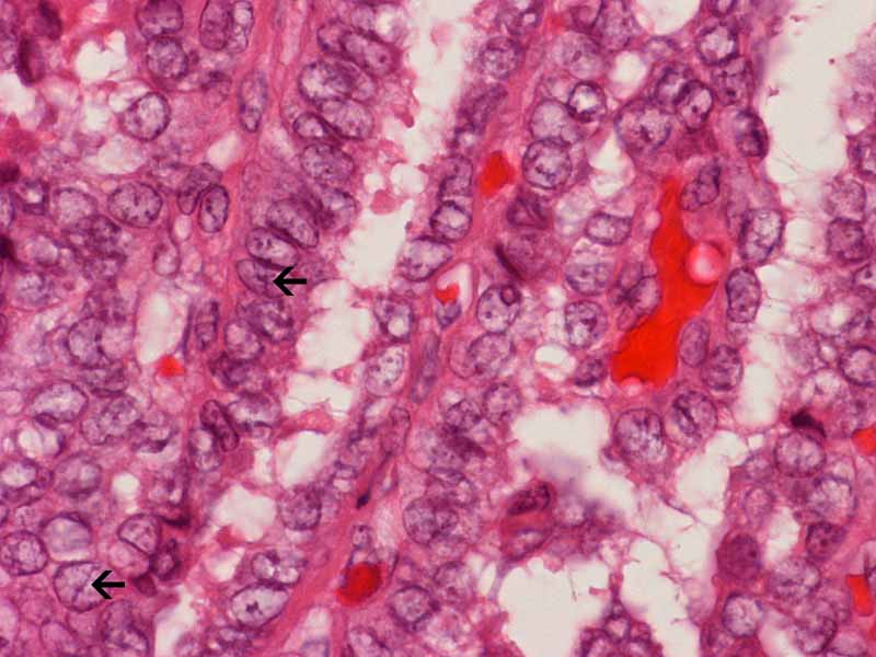
Nuclear features: These high-magnification photos illustrate the characteristic nuclear features of papillary carcinoma of thyroid which include enlarged nuclei with pale "watery" nucleoplasm, pseudonuclear inclusion (arrow in the left panel) and nuclear grooves arrow in the right panel). Overlapping of the nuclei is also a characteristic feature. |
Original slide is contributed by Dr. Kar-Ming Fung, University of Oklahoma Health Sciences Center, Oklahoma, U.S.A.