Case No.: T-002
Diagnosis: Hashimoto thyroiditis
Organ: Thyroid
Last Updated: 07/05/2010
|
|
|
Hematoxylin & eosin |
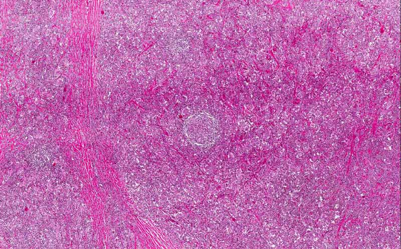
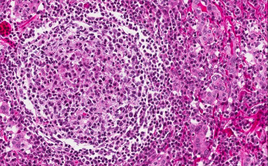
The changes are rather similar in different part of the thyroid. In essence, there is a dense infiltration of mixed population of lymphocytes and plasma cells. Occasional germinal centers are present. |
|
Hematoxylin & eosin |
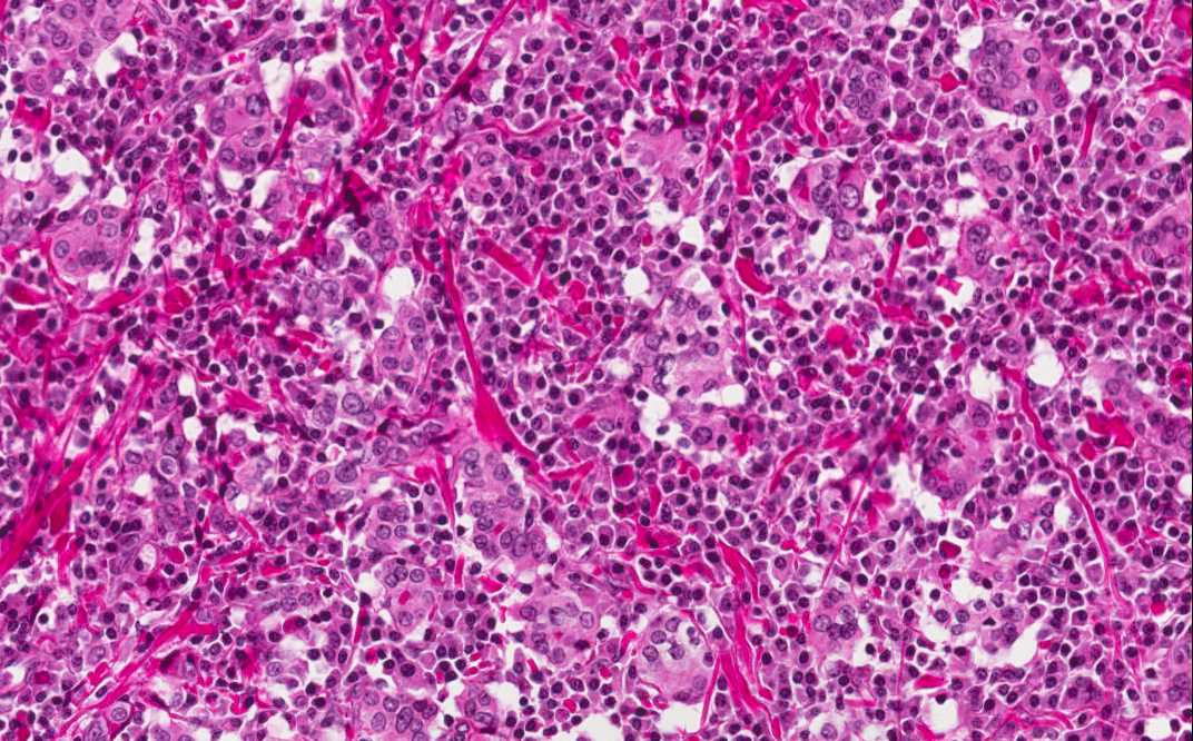
Many small islands of Hürthle are present (arrow) within this background of lymphocytes and plasma cells. |
|
History: The patient was a 55 year-old woman who has a diffusely enlarged thyroid. She was treated with thyroid suppression medication (Synthroid) for two years but the thyroid continued to enlarge despite therapy. Serological studies demonstrated circulating anti-thyroblobulin and anti-thyroid peroxidase antibody. A total thyroidectomy was performed.
Capsule Summary: Hashimoto thyroiditis is a rather common disease. It falls into the spectrum of autoimmune thyroid diseases. The thyroid follicles is gradually destroyed by a variety of cell or antibody mediated immune processes. Patients often have hypothyroidism and episodes of hyperthyroidism. Antibody against the thyroid glands may be present in the blood. Histologically, it is characterized by extensive destruction of thyroid follicles, intense lymphocytic infiltration predominantly of T-cells, lymphoid follicle formation and Hürthle cell formation. Hashimoto thyroidits is a risk factor for the development of non-Hodgkin's lymphoma of the thyroid. For this reason, the specimen must be thoroughly sampled. [more on clinical features of Hashimoto thyroiditis]
Histologic Highlights of this Case:
|