Case No.: U-001  Quiz 

Diagnosis: Atypical mucin secreting glands in pituitary gland

Organ: Pituitary gland

Last Updated: 12/21/2010


Online Slide/Full Screen/Open with ImageScope

Hematoxylin & eosin

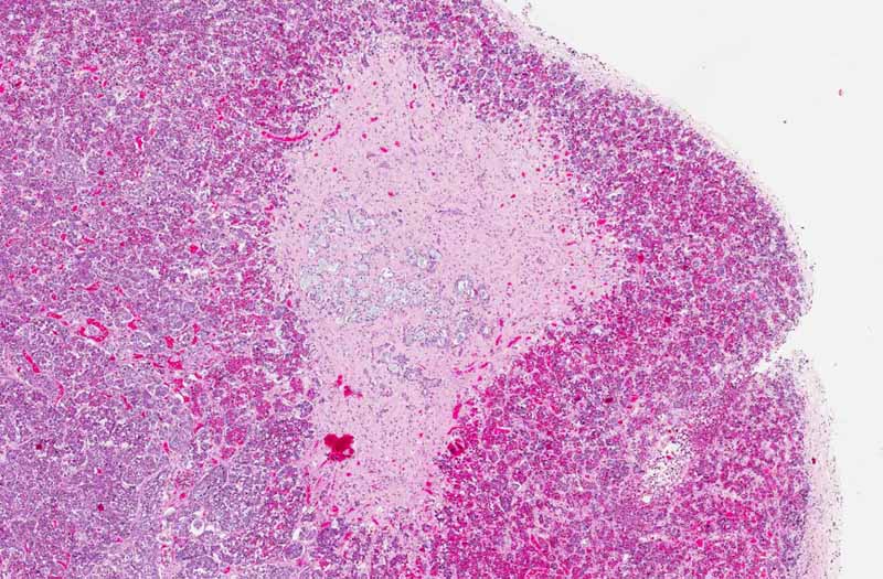
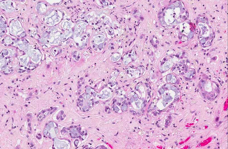
Area 1: Note that within the pale area are clusters of mucin producing glands with basally located nuclei associated with fibrosis.This location is not the most typical location of these glands. Fibrosis should not be associated with ectopic salivary glands.

Hematoxylin & eosin

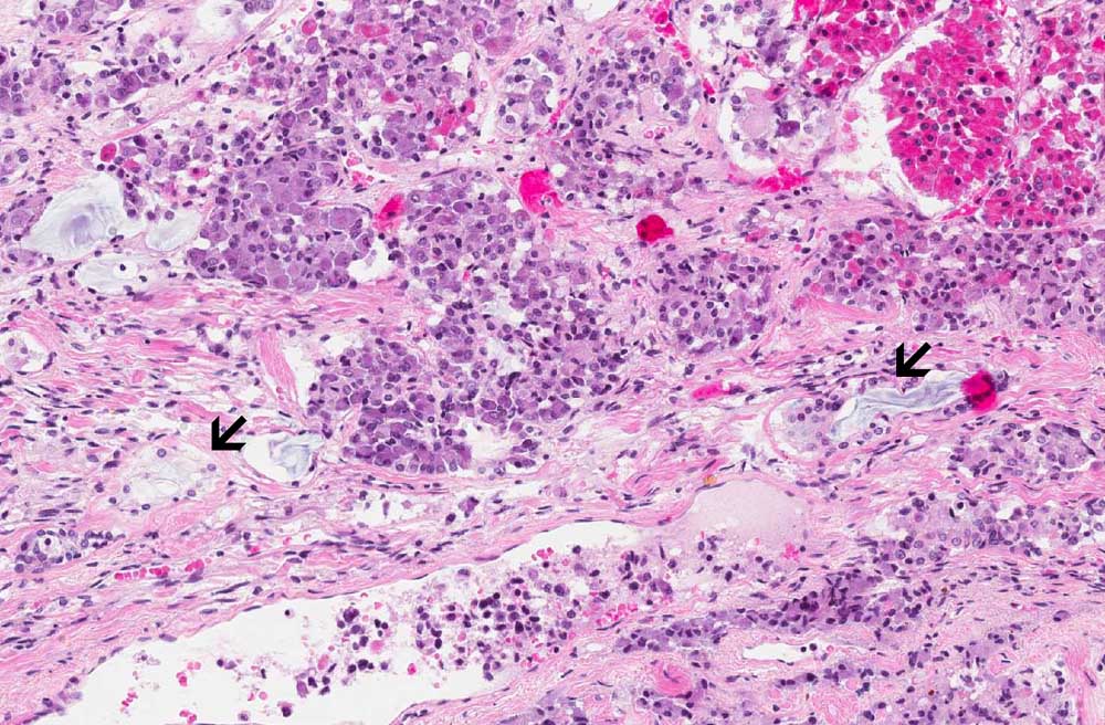
Area 2: Note that less prominent mucin producing glands are found at the interface between the neurohypophysis and adenophysis, a more typical location for these ectopic glands.

History: The patient was a 53 year-old man who had inflammatory bowel disease and peri-rectal abscess which required drainage. He developed sepsis and was hospitalized. A second drainage was performed in addition to treatment with antibiotics. He died unexpectedly of pulmonary embolism. At autopsy, there was no significant gross or microscopic pathologic findings in the central nervous system. Although the pituitary was grossly unremarkable, some interesting findings were found under the microscope as illustrated here.


Histologic Highlights of this Case:

  • The image being shown here is largely composed of the anterior pituitary. Portion of the posterior pituitary is also present. The nests of epithelial glands in the anterior are rather monotonous in size and there is no evidence of hyperplasia or adenoma formation. Second, they are associated with a substantial amount of fibrosis. This raises a possibility of metastatic adenocarcinoma but no cancer is found on autopsy.

  • There is a triangular pale area with mucin producing glands. These glands are most consistent suggestive of ectopic salivary glands. However, there is fibrosis associated with these glands and the glands also look atypical.

Comment:

  • The true idendity of these glands are difficult to be elucidated. Although they may represent ectopic salvary glands in the pituitary, the location is unusual as they tend to be found at the interface between anterior and posterior pituitary.

  • Ectopic salivary glands should not be mistaken as metastatic carcinoma. These glands are typically found on the surface of the pituitary or in the neurohypophysis, often at the interface between the neurohypophysis (posterior pituitary) and adenohypophysis (anterior pituitary) just posterior to the pars intermedia. They are composed of single layer of columnar or cuboidal epithelium with finely granular cytoplasm that is PAS positive. Oncocytic changes is also often. Mucin producing cells can also be present as illustrated in this case. What is unusual in this case is that there is also fibrosis around these glands.

Original slide is contributed by Dr. Kar-Ming Fung, University of Oklahoma Health Sciences Center, Oklahoma, U.S.A.

 Home Page