Answer and Discussion of Quiz Set: W-003
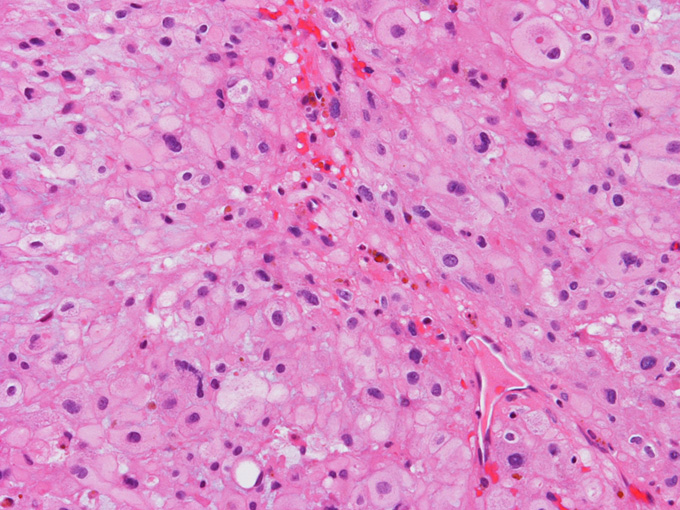
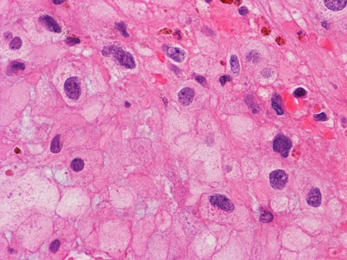
3. The patient is a 65 year-old man who complained of back pain. On his work up, a 5 cm osteolytic lesion is noted at the sacrum with a midline location and the tumor is roughly symmetrical. The patient has a history of renal cell carcinoma that was removed about 10 years ago. Histoloigic examination of the resected tumor yielded the following images. Which of the following sets of immunohistochemistry and/or molecular testing would allow you to correctly diagnose this tumor?
A. CD1a, CD163, S100, cytokeratin AE1/AE3.
B. Isocitrate dehydrogenase 1 and 2 gene (IDH2, IDH2), BRAF mutation and fusion, isochromosome 17.
C. Pax8, CD10, cytokeratin AE1/AE3.
D. Synaptophysin, S100, neurofilament.
E. S100, brachyury, CD10, cytokeratin AE1/AE3.
Answer and Discussion: The answer is (E). By histologic features, this is a chordoma. The image being shown here is classic for this tumor. Although the fine vacuoles of physaliphorous cell in chordoma is not always obvious, the ones in these images are classics. Chordoma is positive for cytokeratin and brachyury and variably positive for S100. As the patient has a history of renal cell carcinoma, metastatic renal cell carcinoma should be ruled out. Renal cell carcinoma is positive for both cytokeratin and CD10. The third possibility is a chondrosarcoma. These tumors, in contrast to chordoma that almost always located right at the midline, has a tendency to deviate to one side. Chondrosarcoma is active for S100 but negative for cytokeratin and brachury.