Case No.: Z-002
Diagnosis: Merkel Cell Carcinoma
Organ: Skin (Face)
Last Updated: 08/28/2010
|
|
|
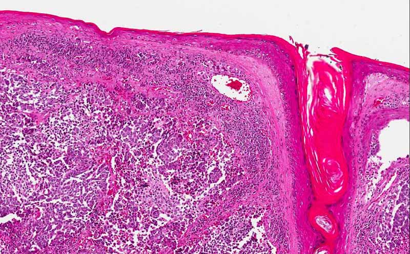
Hematoxylin & eosin |
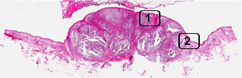
Area 1: Tumor involvement is limited to the dermis without infiltration into the epidermis. In fact, the tumor is separated from the dermis by a thin layer of fibrous tissue. On high magnification, the cells have hyperchromic nuclei, no prominent nucleoli, and only a small amount of cytoplasm. All of these are features of a so-called small blue cell tumor. |
|
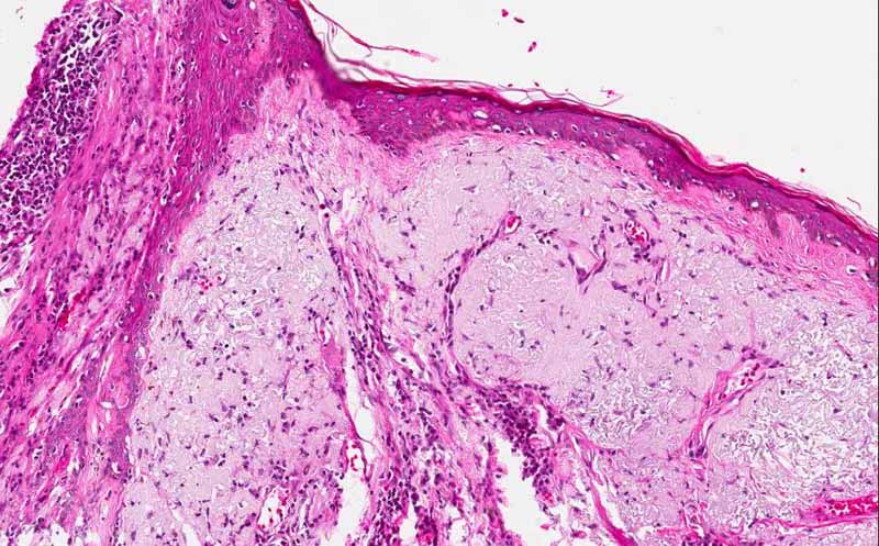
Hematoxylin & eosin |
Area 2: This is taken at the junction between the tumor and the adjacent skin uninvolved by the carcinoma. There is a thick layer of bluish mucoid appearing material in the dermis of the uninvolved skin. These changes are what is termed solar elastosis and is indicative of sun damage. It is typically seen in sun exposed skin. |
|
History: The patient is a 49 year old man who had a roughly 1 cm nodule on his left face. The epidermis covering the nodule appeared to be intact. The lesion was excised along with radical neck dissection of his left neck. The slide illustrated here was obtained from the lesion on his face. No lesion is noted in his lung or other part of the body.
Histopathology:
Comment:
Reference:
|
Bonus Images:
|
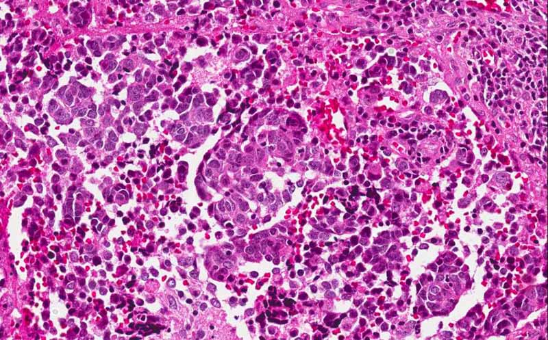
Hematoxylin & eosin |
This image was taken at 60x original magnification. The features of a small blue cell tumor is well illustrated here. |
|
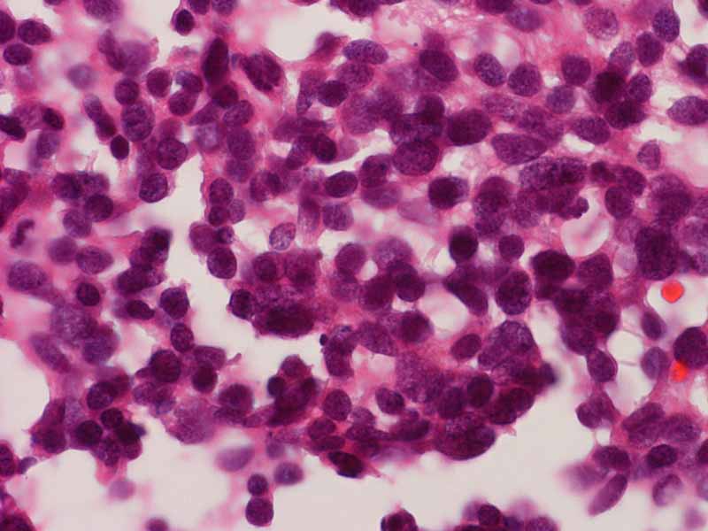
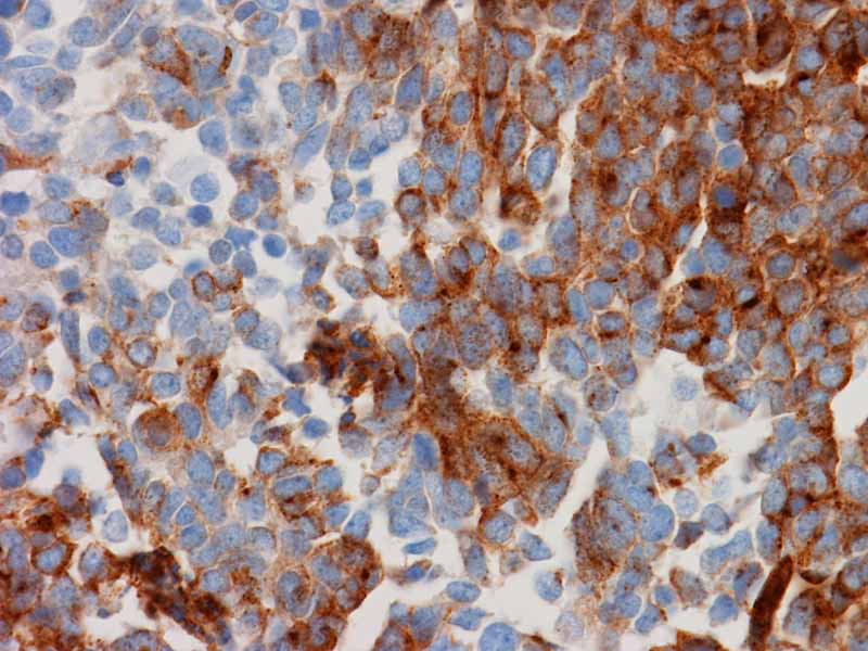
Synaptophysin |
Merkel cell carcinoma is a neuroendocrine neoplasm and is positive for synaptophysin and chromogranin (not shown here). |
|
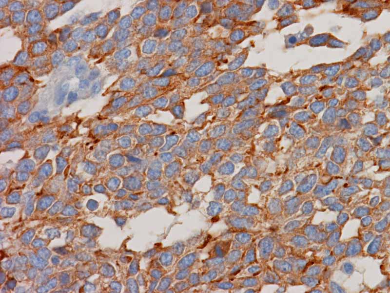
Epithelial Membrane Antigen (EMA) |
Merkel cell carcinoma is also positive for epithelial markers including cytokeratin and EMA. |
Original slide is contributed by Dr. Kar-Ming Fung, University of Oklahoma Health Sciences Center, Oklahoma, U.S.A.