Case No.: Z-004
Diagnosis: Pilonidal cyst
Organ: Skin, buttock
Last Updated: 12/21/2011
|
|
|
Hematoxylin & eosin |
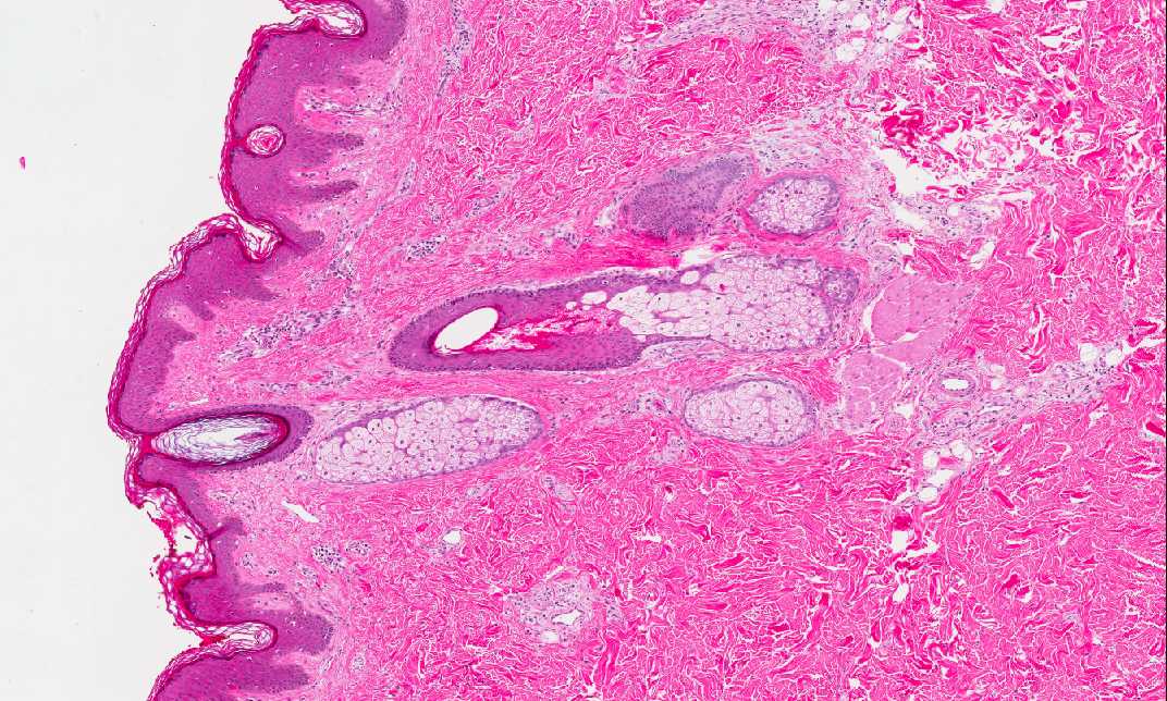
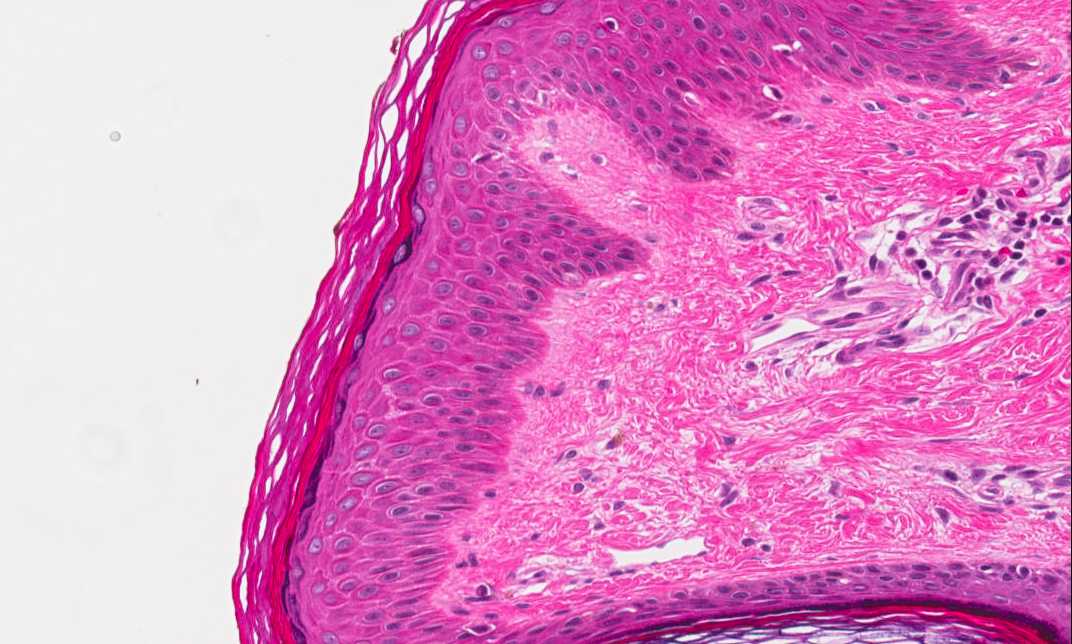
Area 1: This image is taken from the epidermis. You can see keratinizing squamous epithelium with skin appendages. These features identify this specimen as skin. |
|
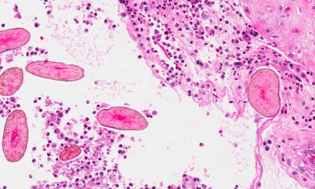
Hematoxylin & eosin |
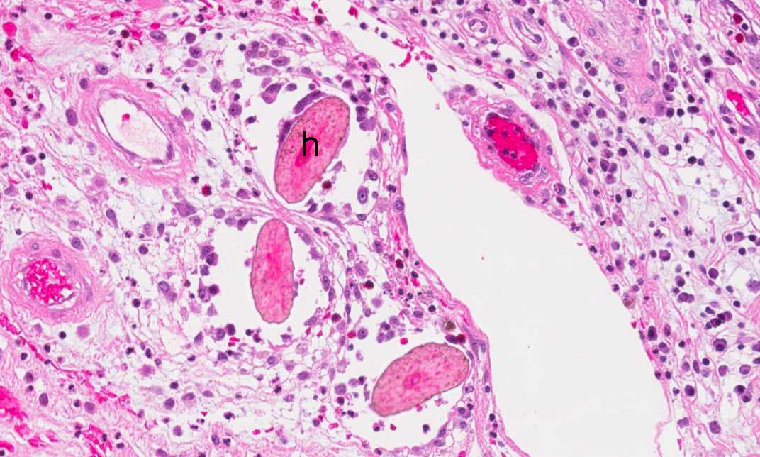
Area 2: The periphery of the lesion is lined by granulation tissue, chronic inflammatory cells and many oval structures. These structures are oblique cuts of hair shafts (h). The granulation tissue and chronic inflammation are reactive changes. |
|
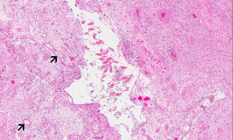
Hematoxylin & eosin |
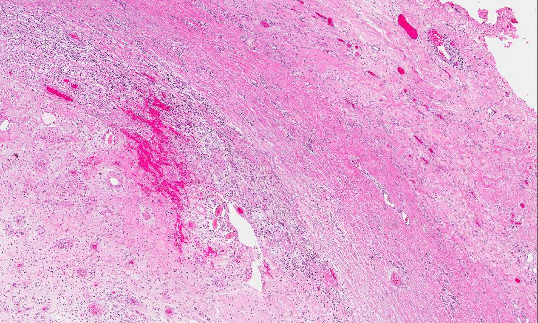
Area 3: Note that the center of the cystic lesion is also filled by a few hair shafts. The granulation tissue and inflammatory cells are reactive changes. Note that numerous blood vessels (arrows) are present in the granulation tissue. |
|
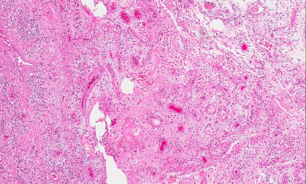
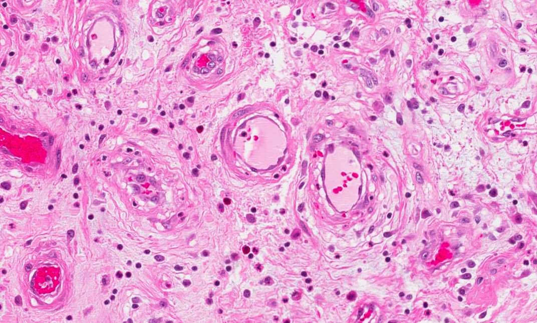
Hematoxylin & eosin |
Area 4: The pathologic changes are very similar in different parts of the cystic lining. |
|
History: This slide was obtained from a nodule, 1.8 cm across, of a 34 year-old woman. The nodule is located at the cleft of the buttock a few centimeters from the anus.
Histologic Highlights of this Case:
Comment:
|
Original slide is contributed by Dr. Kar-Ming Fung, University of Oklahoma Health Science Center, Oklahoma, U.S.A.