| A 55 year-old Man with a Rapidly Enlarging
Thyroid Mass. April, 2003, Case 304-1. Home Page |
Cheng Z. Liu, M.D., Ph.D. and Kar-Ming Fung, M.D., Ph.D. Last update: April 30, 2003.
Department of Pathology, University of Oklahoma Health Sciences Center, Oklahoma City, Oklahoma
Clinical information: 55 year-old man with a rapidly enlarging thyroid mass.
Pathology of the case:
Gross pathology: A total thyroidectomy was performed. A 3 cm fleshy non-encapsulated mass was found on the right lobe with focal extrathyroidal invasion.
Histopathology:
|
|
|
|
|
|
|
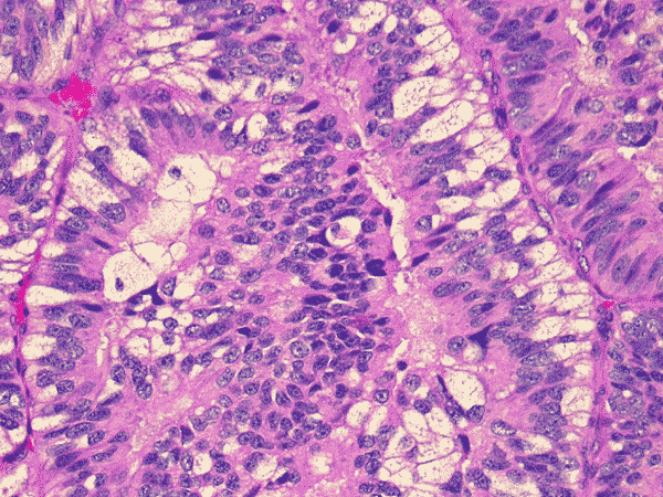
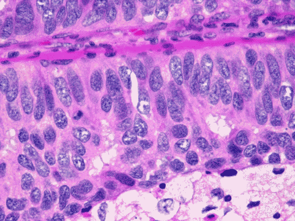
| A. | B. | C. | D. | E. | F. |
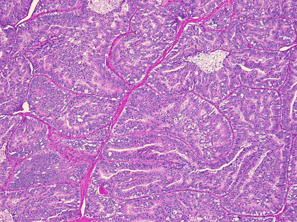
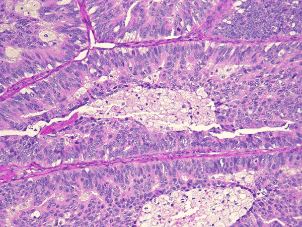
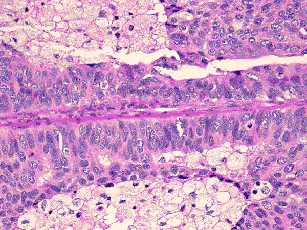
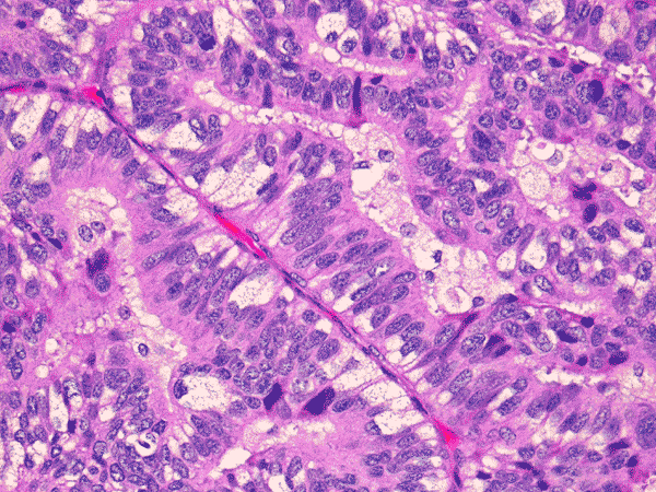
The histologic pictures in different parts of the tumor were very similar. The tumor was composed predominantly of papillary structures covered by tall columnar cells with with nuclear stratification as shown in panel A, B, and C. The tumor nuclei were large, hyperchromatic and elongated; nuclear grooves or ground-glass nuclei typical of papillary carcinoma were not seen. The cytoplasm was granular and often contained subnuclear vacuolizaztion D, E, and F. No solid areas or spindle cell areas were founded. No definitive vascular invasion could be documented.
|
DIAGNOSIS: Columnar cell variant of papillary carcinoma (columnar cell carcinoma) of thyroid. |
Discussion: General Information Pathology Differential diagnosis
General Information
These tumors are aggressive and have high mortality. They tend to occur in a
population that is older that those associated with other papillary carcinoma
and they also have a male predilection. There is a high frequency of distant
metastases, especially to the lung and vertebra, and to regional lymph nodes.
Macroscopically, the tumors are large. They may or may not have a capsule. Extrathyroidal invasion is common. On scanning-magnification, columnar cell carcinoma is characterized by predominantly papillary growth pattern with stratified tumor cells somewhat lined in long paralleled ribbons. Other patterns, such as microfollicular, cribiform, organoid, and solid as well as poorly differentiated areas may be focally evident in some cases. The papillare are lined by tall columnar cells without typical nuclear features seen in those conventional papillary carcinomas. Instead, the nuclei are elongated, euchromatic or hyperchromatic. Nuclear stratification is a typical feature. Mitotic figures are often numerous. Cytoplasm is often scant, and when present, it is usually clear or granular with subneclear vacuolization. In fact, the general cytologic features of the columnar cell carcinoma resemble those seen in early secretory endometrium or endometrioid carcinoma. Areas with usual nuclear features of papillary as well as psammoma body can be focally present. They also possess immunorectivity for thyroidglobulin. It is often helpful to exclude the possibility of a metastatic endometrioid carcinoma or colorectal carcinoma by demonstrating immunoreactivity for thyroidglobulin in these tumors.
Differential diagnosis
The histologic appearance of
columnar cell carcinoma is quite distinct and a correct diagnosis should be
rendered if most of distinctive features are observed. The lesion should not be
confused with the tall cell variant of papillary carcinoma. It is present more
often in women. The tall cells are defined as cells with the height two times
that of the width. The tumor cells have prominent and plentiful eosinophilic and
finely granular cytoplasm due to accumulation of mitochondria; the cytoplasmic
membrane is sharply delineated. Occasional
subnuclear vacuolization may be present. The nuclei in tall cell variant are
enlarged, with nuclear pseudoinclusion and groove; they are situated in the
center or basilar portion of the cell without nuclear stratification. Despite
these differences between the two tumors, tumors with mixed features of columnar
cell and tall cell have been reported
1.
Thyroid tumors with columnar features were first described by Evans 2. Some investigators 3, 4 recognize these tumors as a separately entity as columnar cell carcinoma. Others recognize them as a variant and named them columnar variant of papillary carcinoma 5. The importance of histologic grade over architecture as a prognostic index 6 has also been raised and columnar cell carcinomas are regarded as high-grade histologically and poor prognosis. There is no question that these are aggressive tumor but the name still has to be settled.
Reference:
Akslen
LA, Varhaug JE. Thyroid
carcinoma with mixed tall-cell and columnar-cell features.Am J Clin Pathol. 1990;94:442-5.
Evans
HL. Columnar-cell
carcinoma of the thyroid. A report of two cases of an aggressive variant of
thyroid carcinoma. Am J Clin Pathol. 1986;85:77-80.
Hui
PK, Chan JK, Cheung PS, Gwi E. Columnar
cell carcinoma of the thyroid. Fine needle aspiration findings in a case.
Acta
Cytol. 1990;34:355-8.
Chan,
JKC. Tumors of the thyroid and parathyroid glands in Diagnostic
Histopathology of Tumors, edited by Fletcher CDM. Church Livingstone,
2000.
Wenig
BM, Thompson LD, Adair CF, Shmookler B, Heffess CS.Thyroid
papillary carcinoma of columnar cell type: a clinicopathologic study of 16
cases. Cancer. 1998;82:740-53
Cases of the Month Evaluation Coordinator: KarMing-Fung@ouhsc.edu