| A 45 year-old Woman with an Enhancing Brain
Mass. June, 2003, Case 306-1. Home Page |
Glen D. Houston, D.D.S. and Kar-Ming Fung, M.D., Ph.D. Last update: November 15, 2006.
Department of Pathology, University of Oklahoma Health Science Center, Oklahoma City, Oklahoma.
Clinical information: The patient was a 21 year-old white male who presented with a painless, expansile, and radiolucent mass in the left, posterior mandible for an unknown period of time. A biopsy was performed and yielded the following material.
|
|
|
|
|
| A. | B. | C. | D. |
|
|
|
|
|
| E. | F. | G. | H. |
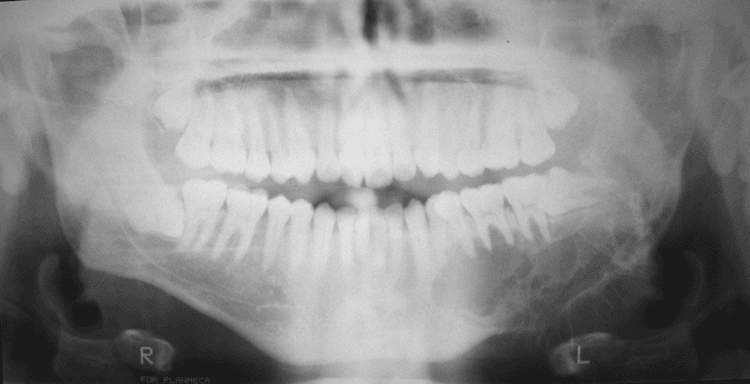
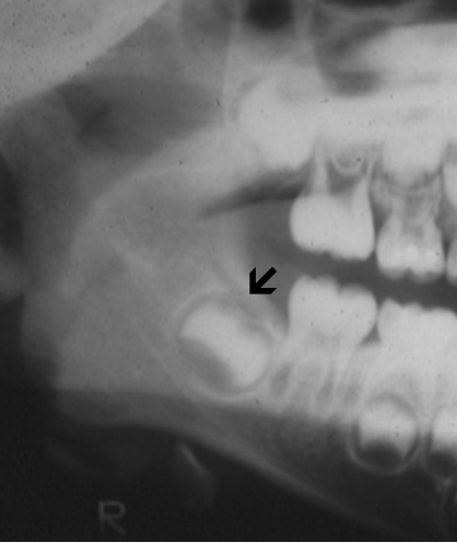
Radiology of the case:
Panoramic radiography shows a radiolucent, multilocular mass that expanded the left ramus (A). It involves the ramus tooth #17 to #21 and also extends into the left condyle. The margin of the lesion is scalloped and non-sclerotic. No periosteal reaction is present. Some fine trabeculae are present but no radiodensity suggestive of mineralized content are present.
Pathology of the case:
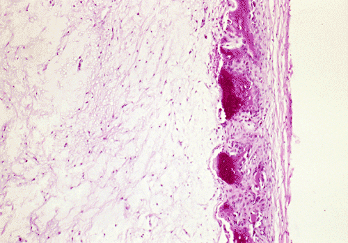
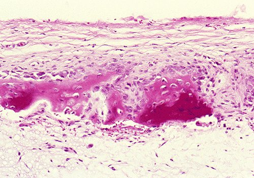
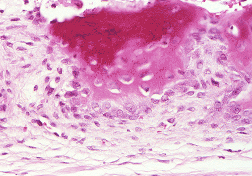
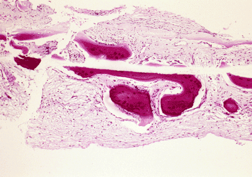
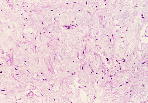
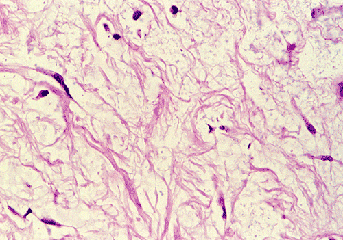
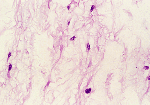
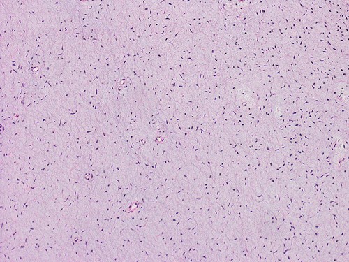
The biopsy material contained several small cores of tissue. Panel B is a low-magnification photo that showed a myxomatous lesion on one side with some linear mature bone trabeculae on the other side. This area probably represented the thinned out residual cortical bone. Panels C and D are higher magnification photos of the bone trabeculae in Panel B. No osteoclastic activity is evident. Panel E shows small, mature bone spicules that are surrounded by myxomatous tissue. The hypocellular and myxomatous nature of the lesion is well demonstrated in Panel F. Panel G and H are high magnification photos from different areas and show a modest number of collagen fibers within a myxomatous background. The bland features of the tumor cells are also well illustrated.
An en bloc resection was performed. The thin cortex that is demonstrated in the panoramic view is confirmed on histologic examination and illustrated in Panel I.
| DIAGNOSIS: Myxoma of the mandible. |
Discussion: General Information Radiographic features Pathology Differential diagnosis
General Information
Myxomas
of the jaw (MOJs) are benign, slow growing, locally invasive, myxomatous
neoplasms
1.
Local recurrence is common. MOJs are rare tumors
and comprise about 8% of all odontogenic tumors
2.
Although they are seen in a wide age group, they are most often found in the
second and third decades. About 12.5% of them occur in children according to one
study
3.
The earliest stage is typically asymptomatic and detection at this stage is by on routine radiographic examination. The earliest
manifestation of myxoma of the
jaw is typically a painless, asymptomatic expansile mass. Teeth in the
affected may spread, migrate, and exhibit root resorption. MOJs are more common in
the mandible than maxilla
4,5.
Maxillary MOJs can spread rapidly through the cancellous bone and behave
clinically more aggressive than mandibular myxomas; some of these tumors may
involve the maxillary sinus with extension into the soft tissue of the cheek
producing a
subcutaneous mass. On physical examination, they can also be mistaken as tumors
arising from the major salivary glands. Due to their notorious risk of
recurrence, en bloc or marginal resection is the treatment of choice over
curettage but this goal may be difficult to achieve in large maxillary MOJs.
Myxomas
are not seen in the extragnathic skeleton and MOJs are currently considered of
odontogenic origin
6.
Although uncommon, myxomas of the soft tissue are well-recognized
entity in the head and neck region and in other parts of the body. They occur
most commonly in the fourth to seventh decades and most of them arise as
intramuscular tumors [Click here
to see a case of intramuscular myxoma]. They may also occur in the subcutaneous tissues, fascial planes
and in association with neurovascular bundles. The palate and parotid gland are
the most common sites
7.
Radiographic features
The
radiographic features of MOJs are not unique. Typically, they occur as expansile
radiolucent unilocular or multilocular lesions with irregular or scalloped
margin. They are often but not always found in tooth bearing regions
5.
The larger ones tend to be multilocular
4.
A sclerotic margin is not a typical feature. There may be thin trabeculae of
residual bone that are often arranged at right angles to one another within the
radiolucent defect but mineralized tissue is not present. This feature
distinguishes it from many odontogenic lesions such as calcifying odontogenic
tumors and osteoblastomas that contain mineralized tissue. The radiographic
features also overlap with many other agnathic lesions. Large MOJs of the
mandible may have a “soap bubble” appearance that mimic ameloblastomas.
Giant-cell granulomas also have radiological and clinical features similar
to MOJs.
Macroscopically,
MOJs have a gelatinous, loose texture and may be covered by a thin shell of
cortical bone that has been eroded by the tumor. Histologically, they are
typically hypovascular and hypocellular. The lesional tissue is composed of
small, stellate or spindle-shaped cells in a myxomatous background. Nuclei are
usually small and hyperchromatic; mitotic figures are lacking. Scattered small
islands of odontogenic epithelial cells that usually appear inactive are
present
6.
Histochemically, the myxomatous ground substance contains acid
mucoploysaccharide, chiefly hyaluronic acid and chondroitin sulfate. This be
well demonstrated by alcian blue stain. A few delicate collagen fibrils are typically
present. Some cases may have an increased amount of collagen and they are
sometimes termed fibromyxoma. There is no evidence, however, that these tumors
behave differently.
Differential diagnosis
Histologically, several mesenchymal tumors may be confused with MOJs. Mesenchymal tumors with myxoid changes should be distinguished from MOJs. Similar to other osseous lesions, radiographic-pathologic correlation is critical in the diagnosis of particularly when only small biopsy specimen is available as in our case.
Myxoid tissue are present in the
dental
papilla of a developing tooth. Enlarged dental follicle and residual
dental pulp of a tooth that has been extracted may all have histologic features
similar to MOJs. These lesions may be mistaken as MOJs
8.
Dental follicles usually contain odontogenic epithelial cell rests and are often
wholly or partially lined by epithelium; the background is usually fibrous and
has variable degree of myxomatous change. Typically,
the amount of myxoid tissue is minute. Dental pulp
tissues are usually circumscribed nodules with eosinophilic condensation with
increased cellularity at the tissue margin; they also contain islands of
odontogenic epithelium.
 |
 |
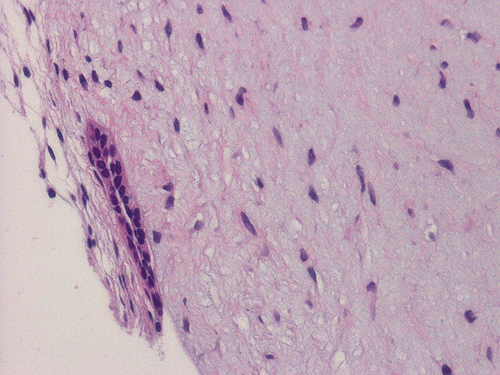 |
| In many cases, the tissue is obtained from the vicinity of a developing tooth as illustrated by the arrow here. | Histologically, the bulk of the tissue appears myxoid, hypocellular, and contains bland, spindle cells. | On careful search, odontogenic epithelium can be found. |
Chondromyxoid fibromas are rare tumors 9,10. They are most commonly seen in patients under 25 years of age. They expand the involved areas and are usually painful. Most of them are found in the mandible. Similar to MOJs, they are circumscribed and radiolucent but it may have mineralized content and a sclerotic margin. Histologically, the myxoid or chondroid components are separated by a more cellular tissue composed of spindle-shaped cells or round cells with varying numbers of multinucleated giant cells. The cells in the chondroid areas usually maintain a spindle-shaped morphology instead of lacunar cells of chondrocytes.
Odontogenic fibroma can occur in any age. Radiographically, it may have a “soap bubble” appearance. Histologically, it is an intraosseous tumor with immature fibrous component with a variable amount of non-neoplastic odontogenic epithelial cell nests. The fibrous component has a homogenous distribution of fibroblasts within a collagenized background. Variable amounts of cementum-like calcifications may be present.
Fibrous dysplasia has demographic features similar to MOJs. Radiographically, it has a “ground-glass” appearance with an ill-defined, hazy margin. Histologically, there are C-shaped trabeculae of woven or immature bone within a proliferating fibroblastic stroma of variable cellularity; myxomatous changes may be present. Osteoblastic rimming of these trabeculae is usually absent. Bone production can be seen at the periphery of the lesion. Many osteoclastic giant cells are present and they have a tendency to form a cuff around capillaries. Extravasated blood and hemosiderin depositionare characteristic features.
Chondrosarcoma of the
jaw is rare. Radiographically, it often contains radiolucent areas and hazy
radiodensity. Although the well-differentiated chondrosarcoma may have bland
histology and mimic normal cartilage, it has chondroid rather than myxoid
background. Mesenchymal chondrosarcoma exhibits increased incidence in the head and
neck area; the presence of highly pleomorphic areas and hemangiopericytoma-like
cells should allow easy separation from MOJs.
Reference:
Barker
BF. Odontogenic myxoma. Semin
Diagn Pathol. 1999 16:297-301.
Auriol
M, Chomette G, Martino R, Bertrand JC, Guilbert F. Odontogenic
myxoma. A histoenzymologic and ultrastructural study. Apropos of 5 cases. J Biol Buccale. 1986 14:215-22.
Keszler
A, Dominguez FV, Giannunzio G. Myxoma in childhood: an analysis of 10 cases. J
Oral Maxillofac Surg 1995 53:518-21.
Kaffe
I, Naor H, Buchner A. Clinical
and radiological features of odontogenic myxoma of the jaws. Dentomaxillofac
Radiol. 1997 26:299-303.
Slootweg PJ, Wittkampf AR. Myxoma of the jaws. An analysis of 15 cases. J Maxillofac Surg. 1986 14:46-52.
Lombardi
T, Lock C, Samson J, Odell EW. S100,
alpha-smooth muscle actin and cytokeratin 19 immunohistochemistry in
odontogenic and soft tissue myxomas. J
Clin Pathol. 1995 48:759-62.
Tse
JJ, Vander S. The soft tissue
myxoma of the head and neck region--report of a case and literature review. Head
Neck Surg. 1985 7:479-83.
Kim J, Ellis GL. Dental follicular tissue: misinterpretation as odontogenic tumors. J Oral Maxillofac Surg. 1993 5:762-7.