| A 5 year-old Boy with
Tetered Cord Syndrome. September, 2003, Case 309-2. Home Page |
Ildiko Nagy, M.D., and Richard Leech, M.D. Last update: September 30, 2003.
Department of Pathology, University of Oklahoma Health Sciences Center, Oklahoma City, Oklahoma
Clinical information:
The patient was a 5 year-old boy with tethered cord syndrome. Imaging studies revealed a small cystic mass at the filum terminale. The following specimen was obtained after surgical release of the tethered cord.
|
|
|
|
|
|
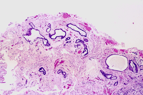
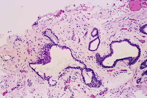
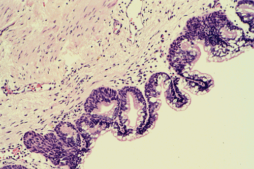
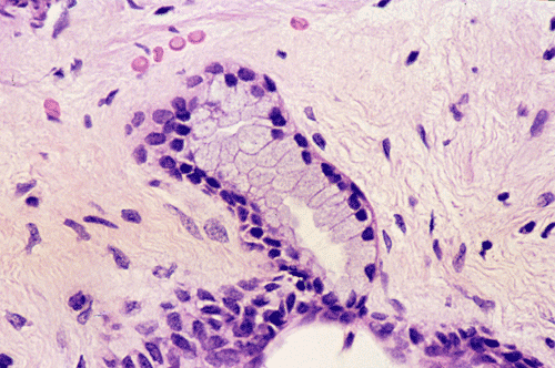
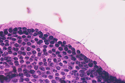
| A. | B. | C. | D. | E. |
Pathology of the case:
On low-magnification (Panel A), the lesion contains has multiple glandular structures in a background of fibroconnective tissue. There is no inflammation. The epithelial component does not seem to have a complex architecture or papilla. On medium-magnification photographs (Panel B and C), the lining epithelium is low-columnar and single layered. Mucin secretion is noted on high-magnification photograph (Panel D). On intraoperative cytologic preparation (squash preparation), the single layer architecture of low-columnar cells is well appreciated (Panel E).
| DIAGNOSIS: Enterogenous cyst of the spinal cord. |
Discussion: What is tethered cord syndrome? General Information Pathology Differential diagnosis Pathogenesis
What is tethered cord syndrome?
A tethered cord refers to a condition in which the lower spinal cord is fixed and restricted from normal ascending movement within the spinal canal. As the spinal column grows, the restricted cord is stretched beyond its tolerance resulting in damage of the nerve roots and blood vessels. Tethered cord syndrome is most commonly seen in children but it can also bee seen in adults 1. Clinically, tethering of the spinal cord results in progressive neurologic impairment as a result of recurrent minor traction injuries. The more common clinical manifestations include pain aggregated by movement, weakness and numbness of the lower extremities and urinary incontinence. Tethered cord syndrome may be associated with other abnormalities such as VATER association. Multiple pathologic conditions can lead to tethering. The most common one is fixation of the spinal cord to a mass of fibroadipose tissue with or without neurological tissue or other tissues. Other conditions include tight filum terminale, secondary adhesion due to inflammation or prior surgery, split cord malformation (diastematomyelia), meningomyelocele, lipomingomyelocele, dermal sinus tract, enterogenous cyst, and teratoma. Dermatologic signs such as hairy patch skin dimple or sinus, capillary hemangioma or atrophic skin may be found in the lumbar region. Early surgical release (detethering) would relieve the symptoms and improve neurologic functions.
General Information on enterogenous cyst
Enterogenous cyst of the
central nervous system, first reported by Pussep in 1934
2,
is a rare congenital anomaly. Subsequent larger series include those reported by
Wilkins and Odum in 1976 3, Wilkins
and Rossitch 1995 4, and Rauzzino
et al. in 2001 5. Enterogenous cysts
can present at a wide range of ages, from the newborn period to the fifth decade
with a slight male predominance (1.8:1)
5.
The spinal cord, especially the lower cervical and upper thoracic regions, is
the most common site. Lumbosacral locations are less frequent. Most commonly,
they are intradural, extramedullary, midline lesion that is ventral to the
spinal cord. However according to a
recent review by Rauzzino, et al., in their series of 13 cases the cysts were
most commonly located dorsal to the cord. A few cases of intracranial location
have also been reported
6,
7,
8,
9.
Enterogenous cysts are often associated with developmental defects of the
overlying skin and/or vertebral bodies. Occasionally they can have fistulous
connection with similar, mediastinal, thoracic or abdominal cysts.
The clinical presentations
of spinal enterogenous cysts are pain localized to the level of spinal cord
affected, myelopathic symptoms, paresis, numbness, or, as in our case, tethered
cord syndrome. The symptoms of spinal cord or nerve root compression may mimic
other space occupying lesions of the spinal canal. Intracranial neurenteric
cysts can be asymptomatic or they can present with neurological symptoms like
ataxia, nystagmus, visual symptoms, cranial nerve palsies that reflect the
structures being compressed or affected.
On non-enhanced CT scan the
lesion appears as a well delineated, irregular, noncalcified, lobulated, cystic
mass that is typically hypodense compared to the adjacent brain or spinal cord
parenchyma. MRI is a better tool than CT for diagnosis. Enterogenous cyst
appears as a sharp-edged lesion. Most of these lesions are iso- to hyper-intense
compared to CSF on T1-weighted images and moderately hyper-intense on proton
density and T2-weighted sequences
10.
Macroscopically the enterogenous cysts are well delineated, thin-walled, fluid-containing masses. The cyst content varies from colorless, transparent fluid resembling CSF to milky or mucinous secretions. Histologically, the cyst is lined by a layer of bland, cuboidal to tall columnar cells with cilia and mucin production. Enterogenous cysts have been classified by Wilkins and Odum 3 into three groups according to histological features. Type A is the simplest type; the walls of these cysts consist of a basement membrane supporting a single or pseudostratified layer of cuboidal to columnar epithelial cells with or without cilia. Type B cysts include more complex elements of the gastrointestinal tract or tracheobronchial tree, including mucus glands and smooth muscle in their wall. Type C cysts have ependymal or glial tissue in addition to the elements seen in the group B cysts. Most enterogenous cysts belong to group A or less frequently to group B.
Differential diagnosis
Enterogenous cysts should be differentiated from other cysts arising in the central nervous system. [Click here to see a table of comparison] It should also be differentiated from choroid plexus tumor and metastatic well differentiated adenocarcinoma. In most cases, it should not be a challenging situation.
A variety of theories have been proposed to explain the occurrence of endodermal tissue in the central nervous system, but none are all inclusive of the different kind of enterogenous cysts. Enterogenous are believed to originate from embryonal dysgenesis. As bone defects are commonly associated with these cysts, it is likely that the primary event occurs in the third week of gestation during gastrulation, when the endodermal and mesenchymal layers are intimately associated with each other. During normal development, this is the time when the primary mesoderm, which gives rise to the notochord, comes to lie in close contact with the endoderm. At a later stage when separation occurs, groups of endodermal cells may be carried back with the mesoderm to give rise to the enterogenous cyst. Alternatively an adhesion between endoderm and ectoderm could cause the "splitting" of notochord 11. The subsequent deficiency in the overlying neural plate could allow for an endodermal diverticulum to herniate and make contact with the surface ectoderm, the so-called “split notochord syndrome”. Immunohistochemical evidence support the notion that enterogenous cyst are derived from primitive gut endoderm 12.
Reference:
Huttmann
S, Krauss J, Collmann H, Sorensen N, Roosen K.
Surgical
management of tethered spinal cord in adults: report of 54 cases.
J
Neurosurg. 2001; 95(2 Suppl):173-8.
Pussep M.
Variete rare de teratome sous-dural de la region cervicale (intestinoma).
Quadriplegie. Extirpation. Guerisan complete. Rev Neurol 1934; 2:879-86.
Wilkins RH,
Odum GL. Spinal intradural cysts. In Handbook of Clinical Neurology, Vol 20. 1976, pp 55-102.
Wilkins
RH, Rossitch JR. Intraspinal cysts.
In Disorders of the pediatric spine. New York: Raven Press, 1995, pp
445-466.
Rauzzino MJ,
Tubbs RS, Alexander E, Grabb PA, Oakes, WJ. Spinal neurenteric cysts and
their relation to more common aspects of occult spinal dysraphism. Neurosurg.
Focus 10: Article 2, 2001.
Ray
A, Chakraborty A, Donaldson-Hugh M.
Enterogenous
cyst of the posterior fossa. Br J Neurosurg. 2000; 14:249-51.
Abe K, Oyama K, Mori K, Ishimaru S, Eguchi M, Maeda M. Neurenteric cyst of the craniocervical junction--case report. Neurol Med Chir (Tokyo). 1999; 39:875-80.
Suri
VS, Tatke M, Sinha S.
Intracranial neurenteric cysts: a report of two cases. Br J Neurosurg.
2002;
16:185-8.
Dias
MS.
Enterogenous cyst of the fourth ventricle: case report. Neurosurgery. 1998;
43:984.
Brooks
BS, Duvall ER, el Gammal T, Garcia JH, Gupta KL, Kapila A.
Neuroimaging features of neurenteric cysts: analysis of nine cases and
review of the literature. AJNR Am J Neuroradiol. 1993; 14:735-46.
Bentley
JF, Smith JR.
Developmental posterior enteric remnants and spinal malformations: the split
notochord syndrome. Arch Dis Child. 1960; 35:76-86.
Mackenzie
IR, Gilbert JJ. Cysts
of the neuraxis of endodermal origin.
J
Neurol Neurosurg Psychiatry. 1991; 54:572-5.