DiffQuick
DiffQuick
DiffQuick
PAP stain
PAP stain
| An 18 year-old Woman with
a Large Mediastinal Mass. October, 2003, Case 310-3. Home Page |
Koushan Siami-Namini, M.D., Jian T. Yang, M.D. Ph.D., Rosemary E. Zuna, M.D., Kar-Ming Fung, M.D., Ph.D. Last update: October 30, 2003.
Department of Pathology, University of Oklahoma Health Sciences Center, Oklahoma City, Oklahoma
Clinical information:
The patient was an 18 year-old man who presented with chest pain and shortness of breath. The pain would last for seconds followed by spontaneous resolution. There was no history of cough, hemoptysis, fever, or chills. A chest x-ray revealed fullness of mediastinum. CT scan revealed a 7.5 x 6.2 x 9.1 cm mass that extended inferiorly along the left superior aspect of the heart. The mass encroached the main pulmonary trunk. Remnant of tissue consistent with residual thymus were noted. The lung was well areated and free of nodules or consolidation. A CT guided fine needle aspiration (FNA) was performed.
A surgery was performed and yielded a 9.5 x 6.5 x 5.5 cm encapsulated mass with attached soft tissue that appeared to be thymic tissue on subsequent microscopic examination. The mass had a gray-yellow, solid cut surface with multifocal hemorrhage and necrosis. The capsule was largely intact with no macroscopic evidence of penetration identified.
Pathology of the case:
|
|
|
|
|
|
|
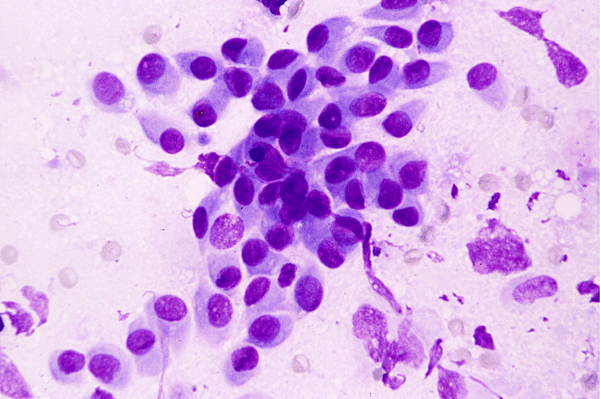
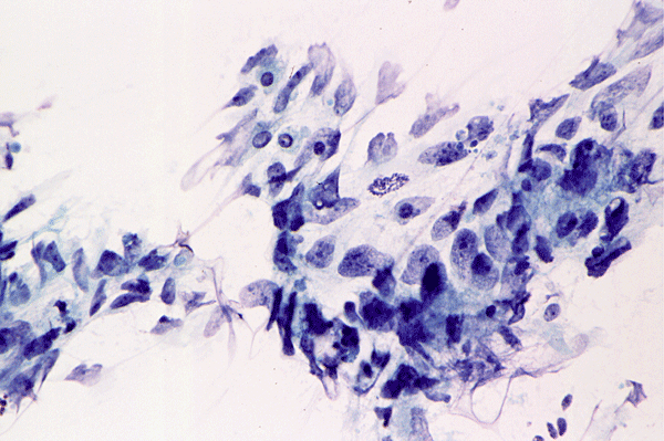
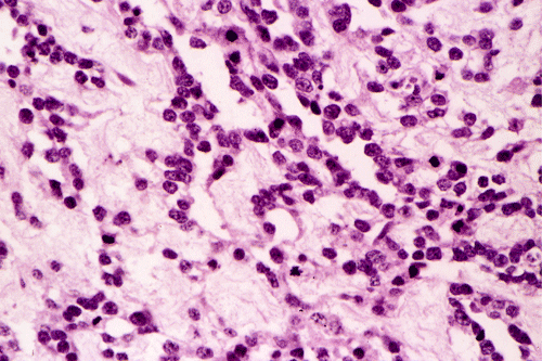
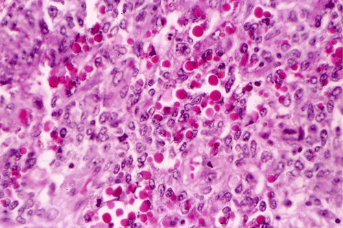
A. DiffQuick |
B. DiffQuick |
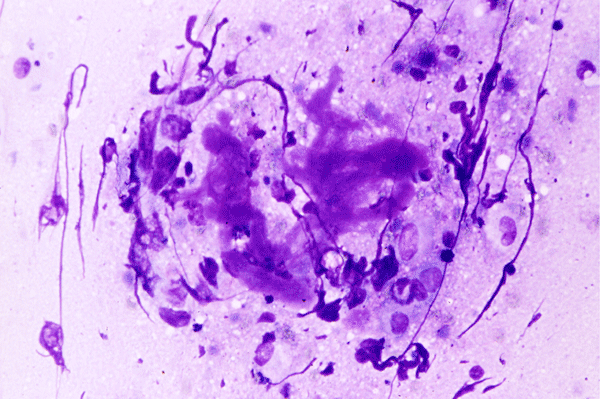
C. DiffQuick |
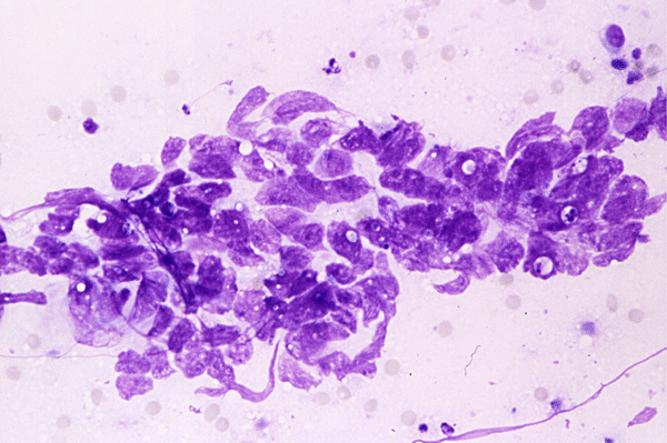
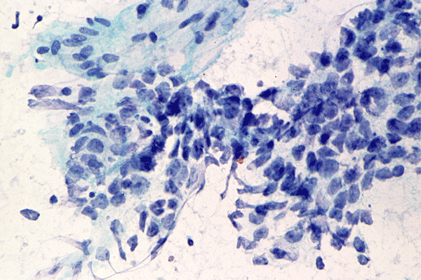
D. PAP stain |
E. PAP stain |
 |
 |
 |
 |
 |
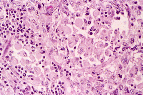 |
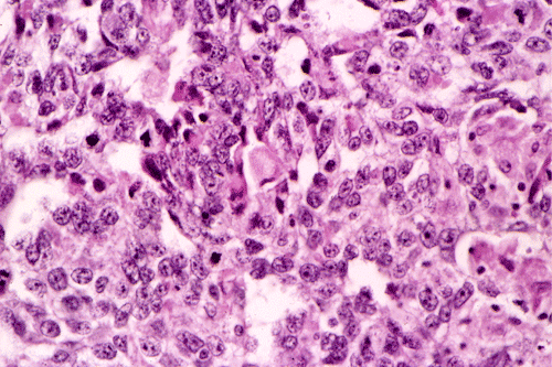
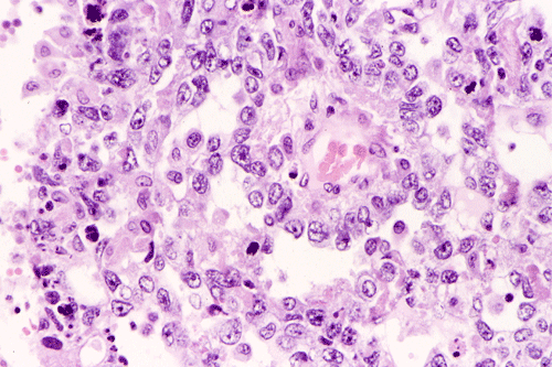
| F. | G. | H. | I. | J. | K. |
 |
 |
 |
 |
||
| L. |
M. PAS |
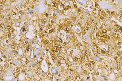
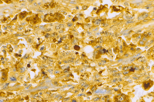
N. a-fetal protein |
O. a1-antitrypsin |
Cytopathology of CT guided FNA specimen:
The fine needle aspiration (FNA) specimen (Panel A, B, C, D, and E) contain clusters of loosely packed cohesive cells without associated lymphoid cells or tigroid background. Some of the cell clusters (Panel A) appear bland but other cells are highly pleomorphic (Panel B, D, and E). The bland appearing cells may represent reactive mesothelial cells and the highly pleomorphic cells are tumor cells. The tumor cells have an epithelioid appearance with large nuclei and moderate amount of cytoplasm. While nucleoli are not prominent in the sections stained by DiffQuick (Panel A and B) , large nucleoli are noted in sections stained by Papanicolaou stain (Panel C, D, and E). Small clumps of acellular hyaline material are present and their metachromatic property is best demonstrate in the DiffQuick stained sections (Panel C). These material appears as bluish green substance on sections stained by Papanicolaou stain. These hyaline material provides good clue for the cytologic diagnosis of a mediastinal yolk sac tumor and have been described by Yang et al. 19, 20. They correspond to the hyalinized eosinophilic globules in yolk sac tumors. Based on these cytologic features, a diagnosis of germ cell tumor with yolk sac tumor component was made.
Gross pathology:
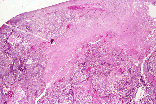
The surgically excised specimen is an encapsulated mass (9.5 x 6.5 x 5.5 cm) attached to a portion of thymus (6.5 x 4.0 x 1.0 cm). The capsule of the mass is 0.1 to 0.3 cm in thickness, smooth and grossly intact. The mass has a gray-brown, and soft cut surface with multifocal hemorrhage. The mass is well contained and dose not invade into surrounding mediastinal structures. The thymus has a tan-yellow, lobulated cut surface withougt any gross abnormality.
Histopathology of surgical specimen:
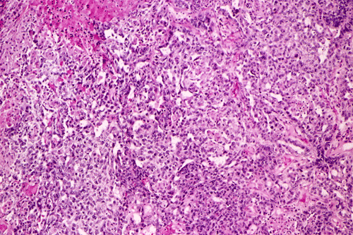
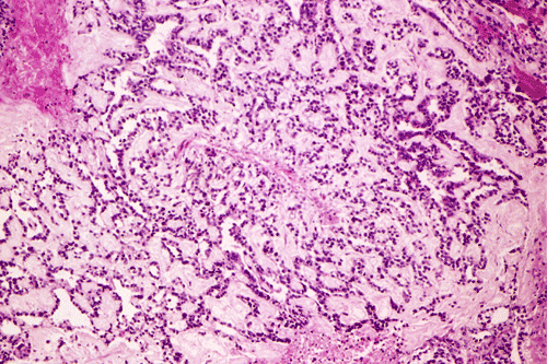
A survey at low-magnification shows a necrotic neoplasm (Panel F). The tumor cells arrange in different patterns. The tumor cells are densely packed in some areas but loosely packed in other areas. At medium-magnification, several different patterns of architecture are disclosed. The most commonly encountered pattern in this case is a reticular-microcystic arrangement of cells (Panel G). In a significant amount of other areas, there are hypocellular to myxoid non-neoplastic stroma lined by a single layer of neoplastic cells (Panel H, I). The tumor cells have moderate to large nuclei with substantial pleomorphism. The chromatin is clumpy and hyperchromatic (Panel J). In some areas the tumor cells has an hepatoid appearance featured by a smaller nuclei with substantial amount of amphophilic cytoplasm (Panel K). Schiller-Duval bodies are occasionally noted (Panel L). A variable number of eosinophilic globules are also present and they are strongly positive for periodic acid-schiff (PAS) reaction (Panel M). Results of immunohistochemistry are as follows:
| Antigen | Result |
| Cytokeratin AE1/AE3 | Strongly positive in practically all tumor cells. |
| a-fetal protein (AFP) | Strongly positive in practically all tumor cells, negative in hyaline globules. |
| a1-antitrypsin (A1-AT) | Strongly positive in practically all tumor cells, positive in hyaline globules. |
| Human chorionic gonadotrophin (hCG) | Rare positive tumor cells. |
| Placental alkaline antigen (PLAP) | Negative in tumor cells. |
| CD30 (Ki-1) | Negative in tumor cells. |
| Epithelial membrane antigen (EMA) | Negative in tumor cells. |
| DIAGNOSIS: Yolk sac tumor (endodermal sinus tumor) of the mediastinum. |
Comment:
Yolk sac tumor arising in the thymus often have extensive invasion into the adjacent tissue. In this cases, the tumor is still confined. The histology is quite typical for a yolk-sac (endodermal sinus) tumor. The reticular-microcystic pattern being illustrated here is the most common pattern being encountered. Only a small number of Schiller-Duval bodies are present. The large cells with hepatoid look correspond to the hepatoid pattern in yolk sac tumor. The solidly arranged sheets of tumor raise the possibility of an embryonal carcinoma. However, the nuclear pleomorphism, although significant, is still short of that from embryonal carcinoma. The extracellular, PAS(+), eosinophilic hyaline globules are also typical for yolk sac tumor. These globules are often immunoreactive for a-fetal protein and a-1-antitrypsin. In our case, they are only positive for a-1-antitrypsin. The lack of CD30 immunoreactivity, again, does not support a diagnosis of embryonal carcinoma.
Discussion: General Information Embryonic Consideration Pathology Differential diagnosis Cytogenetics
General Information
Yolk sac tumor, also known as endodermal sinus tumor, belongs to the family of germ cell tumor. Gonadal and extragonadal germ cell tumors derive from germ cells and they have very diversified phenotypic features. Differentiation of germ cell tumors can be separated into two major groups: tumor with teratomatous differentiation and non-teratomatous differentiation. Teratomatous differentiation can be composed of mature and/or immature tissue. Within the non-teratomatous differentiation, there are two major subtypes: tumor with seminomatous differentiation resembling primordial germ cells (seminoma and dysgerminoma) and non-seminomatous differentiation. The non-seminomatous differentiation occur in the form of undifferentiated component (embryonal carcinoma) or extraembryonic differentiation (yolk sac tumor and choriocarcinoma). All the aforementioned types of differentiations can occur as pure or combined form that lead to a rich diversity in histopathologic features. Gonadal and extragonadal germ cell tumors are essentially identical in phenotypic features. However, the relative incidence of different subtypes varies greatly in different anatomical locations and organs. Moran and Suster provided a useful classification of germ-cell tumor of the mediastinum 1. Interested readers should refer to the comprehensive articles by Moran et al. 1, 2, 3.
|
Classification of germ tumors of the mediastinum 1 |
|
Teratomatous
tumors Mature Teratomas (composed of well differentiated, mature elements) Immature teratomas (with the presence of immature mesenchymal or
neuroepithelial elements) Teratomas with additional malignant components
|
Non-teratomatous
tumors Seminomas Yolk sac tumors Embryonal carcinomas Choriocarcinomas Combined non-teratomatous tumors
|
Primary germ cell tumor is an uncommon tumor of the mediastinum and account for
10-15% of all mediastinal tumors
4.
Primary germ cell tumor of the mediastinum occurs most often in young adults in
the 2nd to 4th decade
5,
6
but can also occur in the very young and elderly
1,
7.
Although mediastinal teratomas are as common in males and females, non-teratomatous
mediastinal germ cell tumors occur almost exclusively in male
5,
1.
Rare cases have been described in female patients
8.
Some
of the mediastinal germ cell tumors remain asymptomatic and are found as
incidental findings. Those tumors that manifest produce symptoms and signs
resulted from compression or invasion of local structures such as compression of
the vena cava. Symptoms are non-specific. The most common ones include chest pain, shortness of
breath, weight loss, chills and fever, superior vena cava syndrome. Trichophysis (coughing up of hair), however, is a
rare but diagnostic feature of teratomas. Association of Kleinfelter’s syndrome
9,
sexual precocity
10
and hematologic malignancies
11
with mediastinal germ cell tumors have also been reported. An increased serum level of a-fetal protein and human chorionic gonadotrophin
suggest the presence of non-seminomatous component.
Among non-semionomatous-non-teratomatous germ cell
tumors of the mediastinum, yolk sac tumor is the most common
3.
Mediastinal
yolk sac tumor was first described by Teilman
12
and comprises more than half of all the cases in the series by Moran et al. The
second most common entity is combined germ cell tumor that comprises about
one-fifth of all cases. Choriocarcinoma and embryonal carcinoma are less common
and each comprises about one-tenth of all cases with embryonal cell carcinoma as
the least common one
3. The diagnosis of primary choriocarcinoma in the
mediastinum can be contraversial due to the highly metastatic nature of
choriocarcinoma.
For treatment and
prognostic purposes, mediastinal germ cell tumors are divided into seminomatous
and non-semintomatous group. A tumor should be treated as non-seminomatous type
if it contains both seminoatous and non-seminomatous components. The long-term
survival with treatment in mediastinal seminomas can reach 85%-90%
13,
14 largely due to its radiosensitivity and also 75% of
these tumors are stage I disease at presentation
1. The non-seminomatous mediastinal germ cell tumors
carry worse prognosis than its gonadal counterparts
1,
3,
14
or retroperitoneal primary germ cell tumors
15.
A 5-year survival of 45% in patients with mediastinal non-seminomatous-non-teratomatous
mediastinal germ cell tumor has been achieved in one study
15.
Yolk sac tumors appear to have worse prognosis
16
and choriocarcinoma is uniformly fatal
1.
With their embryonic origin, it would not be surprised to see that germ cell
tumors occur most often in the gonads. However, they are also seen in other
midline locations such as the pineal gland, sellar-suprasellar region,
retroperitoneum, mediastinum and sacrococcygeal region
17.
During the 4th to 6th week of embryonic development, the
primordial germ cells migrate from their original midline location near the
origin of the allantois to the gonadal ridge of the future gonads
18 along the
dorsal mesentery of the hindgut. This process partly explained the occurrence of
germ cell tumor along the midline. It is well known that the primordial germ
cells disseminate widely throughout many tissues and organ during early
embryonic development. The extragonadal primordial germ cells persist most
frequently in the mediastinum and the diencephalopineal region. The reason for
this distribution remains uncertain. This distribution, however, partly explains the
relatively high frequency of extragonadal germ cell tumor in the mediastinum and pineal region relative
to other midline locations.
Mediastinal non-teratomatous germ cell tumor almost always presents as an
anterior mediastinal mass 3.
Macroscopically, mediastinal germ cell tumor is a large and soft mass with
the size of ranging from 6 to 20 cm in greatest dimension. The cut surface is
homogeneous, freshy, and decorated by areas of hemorrhage and necrosis. They are
unencapsulated and have inconspicuous boundary. Invasion of the surrounding
mediastinal structures is a frequent intraoperative finding. In uncommon cases,
mediastinal seminoma may be accompanied by extensive fibrosis. Histologically,
mediastinal germ cell tumors have features similar, if not identical,
to their gonadal counterparts. Here, we will restrict our discussion to yolk sac
tumors.
Yolk sac tumors exhibit a wide range of histologic patterns that differ
considerably from each other. In most cases, a mixed histologic pattern is
present and, not infrequently, one or two patterns may predominate. The most
common pattern is the microcystic-reticular pattern. The other histologic patterns
included endodermal sinus, solid, alveolar-glandular, polyvesicular vitelline,
myxomatous, macrocystic, papillary, intestinal, hepatoid, and spindle cell.
Although there is a rich variation in architecture, the variations in cytologic
features are less impressive. The tumor cells are medium to large and have clear to pale,
granular cytoplasm. Hobnail cells are not uncommon and often seen with the
papillary pattern. The nuclei are large, vesicular, and contain prominent
nucleoli. Yolk sac tumors are mitotically active. Necrosis and hemorrhage
are common.
Yolk sac tumor often, but not always, contains bright, eosinophilic globules. These extracellular, small, round,
brightly eosinophilic, hyaline, strongly PAS-positive, diastase-resistant globules or droplets are most often associated with the microcystic-reticular pattern and endodermal sinus
pattern. These globules are considered to be secreted by the tumor cells and
accumulate within the cytoplasm. As the amount of secretion increases, the cell
become distended and ruptures, discharging its contents into the surrounding
tissue. Although characteristic, these eosinophilc hyaline globules are not
diagnostic of yolk sac tumor because they are also found in other malignant
tumors particularly those with poor differentiation. When they are present in
specimen from fine needle aspiration, as in our case, they are metachromatic and
provide a good clue for diagnosis
19,
20.
The microcystic-reticular pattern is
characterized by a loose vacuolated network with small cystic spaces or
microcysts that give rise to a meshwork of communicating space. The microcysts
are lined by flat, pleomorphic, epithelium-like cells with large hyperchromatic
or vesicular nuclei. The myxomatous pattern is
similar to the microcystic pattern but is loosely packed and contains mucoid
material.
The papillary or "festoon" pattern is composed of
papillary structures featured by fine fibrovascular cores lined by mitotically
active epithelium-like cells with considerable degree of cellular and nuclear
polymorphism. The connective tissue may show extensive hyalinization.
The
endodermal sinus pattern is
composed of perivascular formations featured by a narrow band of connective
tissue with a capillary blood vessel in the center and lined by a layer of
cuboidal or low columnar embryonal epithelial-like cells. The cells have large,
slightly vesicular nuclei, prominent nucleoli. A single layer of flat cells
lines the surrounding sinusoidal space. These characteristic perivascular
formations, the Schiller-Duval bodies, are considered diagnostic of yolk sac
tumor but they are not always present. Embryologically, the downgrowth of yolk
sac epithelium into the extraembryonic mesenchyme of the placenta in rodent is
known as endodermal sinuses. The Schiller-Duval bodies are similar to the
endodermal sinus of placenta in rodent that was first described by Duval in 1891
21. The loose myxomatous pattern was considered to be analogous to magma
reticulare or the extraembryonic mesoderm of the exocelom. Teilum also
demonstrated that the reticular and papillary patterns recapitulate the
labyrinthine placenta of the rodents
22,
23.
Recognition of Schiller-Duval bodies in fine needle aspiration, although not a
common event, provide good clue for diagnosis
19,
20.
The alveolar-glandular pattern
is composed of alveolar, gland-like, or larger cystic spaces and cavities lined
by flat or cuboidal epithelium-like cells with large, prominent nuclei and
surrounded by myxomatous stroma or cellular aggregates. The
solid pattern is
composed of aggregates of small epithelium-like polygonal cells with clear
cytoplasm and large vesicular nuclei with prominent nucleoli.
Differential diagnosis
Under most situations, the
histopathologic features of yolk sac tumor of the mediastinum allow rapid recognition. The possibility of a metastatic yolk sac tumor must be ruled out
before a diagnosis of primary tumor is made.
Embryonal carcinoma may pose diagnostic problems as it has one or more of
the characteristics of yolk sac tumors. In general, embryonal carcinoma is solid
or glandular in arrangement, has far more pleomorphism and necrosis than yolk
sac tumors. In contrast, yolk sac tumor has clear cells, distinctive histologic
arrangement, and hyaline bodies. Most embryonal cell carcinomas are positive for
CD10. It should also be noted that a small number of yolk sac tumors are also
positive for CD10.
The rare hepatoid and intestinal pattern may suggest metastatic
hepatocellular carcinoma or metastastatic adenocarcinoma. Again, careful search
for the distinctive histologic features of yolk sac tumor as well as
immunohistochemistry would be helpful in establishing the correct diagnosis. The
spindle cell pattern may suggest sarcoma.
Cytogenetics and association with hematologic malignancies
Isochromosome 12p has been demonstrated in gonadal and extragonadal germ
cell tumors
27,
28
which often lead to a gain of sequences. The critical region involved in the
pathogenesis of germ cell tumor appear to be located at chromosome 12p11.1-12p12.1
29
and the genes involved may be associated with suppression of apoptosis and
Sertoli cell-independent of during progression from in situ to invasive
testicular germ cell tumor
30.
Mediastinal germ cell tumor is associated with an increased incidence of
hematologic malignancy with acute megakaryoblastic leukemia and malignant
histiocytosis as the most common associated hematologic malignancy. In the study
performed by Nichols et al.
11,
the median time between the diagnosis of mediastinal germ cell tumor and development of hemopoietic
malignancies was approximately 5 months. It does not seem to be a result of
cisplatin-based chemotherapy in patients with mediastinal germ cell tumor. This phenomenon
is restricted in patients with non-seminomatous mediastinal germ cell tumors
particularly those with histologic or serologic evidence of yolk sac tumors
11,
31,
32,
33. Isochromsome
12p, a rare genetic change in hematopoietic malignancies, has bee identified in
the leukemic component of these cases
33,
34.
These findings suggest that the mediastinal germ cell tumor and simultaneously occurring
hematopoietic malignancy may share the same precursor cells in the mediastinum.
Interestingly, isochromosome 12p has also been identified in two cases of acute
myeloid leukemia that does not have evidence of mediastinal germ cell tumor
35.
This further raises the question on the role of isochromosome 12p in the
pathogenesis of mediastinal germ cell tumorand hemoatologic malignancies.
Reference:
Moran
CA, Suster S.
Primary germ cell tumors of the mediastinum I. Cancer
1997;80:681-90.
Moran
CA, Suster S, Przygodzki RM, Koss MN.
Primary germ cell tumors of the mediastinum II. Cancer
1997;80:691-8
Moran
CA, Suster S, Koss MN.
Primary germ cell tumors of the mediastinum III. Cancer
1997;80:699-707.
Davis
RD Jr, Oldham HN Jr, Sabiston DC Jr.
Primary cysts and neoplasms of the mediastinum: recent changes in clinical
presentation, methods of diagnosis, management, and results. Ann Thorac
Surg.
1987;44:229-37.
Chen
JKC. Tumors of the lymphoreticular system, including spleen and thymus. In:
Fletcher CDM, editor. Diagnostic Histopathology of Tumors. 2nd
edition. London: Churchill Livingstone; 2000, p. 1295-8.
Knapp
RH, Hurt RD, Payne WS, Farrow GM, Lewis BD, Hahn RG, Muhm JR, Earle JD.
Malignant germ cell tumors of the mediastinum. J Thorac Cardiovasc Surg 1985;89:82-9.
Mullen
B, Richardson JD.
Primary anterior mediastinal tumors in children and adults. Ann
Thorac Surg 1986;42:338-45.
Gooneratne
S, Keh P, Sreekanth S, Recant W, Talerman A.
Anterior mediastinal endodermal sinus (yolk sac) tumor in a female infant. Cancer 1985;56:1430-3.
Bebb
GG, Grannis FW Jr, Paz IB, Slovak ML, Chilcote R.
Mediastinal germ cell tumor in a child with precocious puberty and
Klinefelter syndrome. Ann Thorac Surg. 1998;66:547-8.
Schwabe
J, Calaminus G, Vorhoff W, Engelbrecht V, Hauffa BP, Gobel U.
Sexual precocity and recurrent beta-human chorionic gonadotropin upsurges
preceding the diagnosis of a malignant mediastinal germ-cell tumor in a
9-year-old boy. Ann Oncol. 2002;13:975-7.
Nichols
CR, Hoffman R, Einhorn LH, Williams SD, Wheeler LA, Garnick MB.
Hematologic malignancies associated with primary mediastinal
germ-cell tumors. Ann Intern Med. 1985;102:603-9.
Teilmann
I, Kassis H, Pietra G.
Primary germ cell tumor of the anterior mediastinum with features of
endodermal sinus tumor (measoblastoma vitellinum). Acta
Pathol Microbiol Scand 1967;70:267-78.
Hainsworth
JD, Greco FA. Germ cell
neoplasms and other malignancies of the mediastinum. Cancer Treat Res.
2001;105:303-25.
Sham
JS, Fu KH, Choi PH, Lau WH, Khin MA, Choy D.
Primary mediastinal seminoma. Oncology. 1990;47(2):124-7.
Bokemeyer
C, Nichols CR, Droz JP, Schmoll HJ, Horwich A, Gerl A, Fossa SD, Beyer J,
Pont J, Kanz L, Einhorn L, Hartmann JT.
Extragonadal germ cell tumors of the mediastinum and retroperitoneum:
results from an international analysis. J Clin Oncol. 2002
;20:1864-73.
Saxman
S, Nichols CR, Williams SD, Loehrer PJ, Einhorn LH.
Mediastinal yolk sac tumor. The Indiana University experience, 1976 to 1988.
J Thorac Cardiovasc Surg. 1991;102:913-6.
Johnson
DE, Laneri JP, Mountain CF, Luna M.
Extragonadal germ cell tumors. Surgery
1973;73:85-90.
Moore
KL. The developing human. 4th edt. Philadelphia: WB
Saunders, 1988. p 262
Yang
GC, Hwang SJ, Yee HT.
Fine needle aspiration cytology of unusual germ cell tumors of the mediastinum: atypical seminoma and parietal yolk sac tumor. Diagn
Cytopathol 2002;27:69-74.
Yang
GC. Fine needle
aspiration cytology of Schiller-Duval bodies of yolk sac tumor. Diagn
Cytopathol 2000;23:228-32.
Duval
M. Le placenta des rongeurs. J Anat Physiol (Paris) 1891;27: 24–73,
344–395, 513–612.
Teilum
G. Classification
of endodermal sinus tumour (mesoblastoma vitellinum) and so-called
“embryonal carcinoma” of the ovary. Acta Pathol Microbiol Scand 1965;
64:407-429.
Teilum
G. Special tumors of the ovary and testis, and related extragonadal lesions;
comparative pathology and histological identification. Philadelphia: JB
Lippincott., 1976. p.33-115.
Moran
CA, Suster S. Hepatoid
yolk sac tumors of the mediastinum: a clinicopathologic and
immunohistochemical study of four cases. Am
J Surg Pathol. 1997;21:1210-4.
Moran
CA, Suster S. Yolk sac
tumors of the mediastinum with prominent spindle cell features: a
clinicopathologic study of three cases. Am J Surg Pathol. 1997;21:1173-7.
Teilum
G, Albrechtsen R, Norgaard-Pedersen B.
The histogenetic-embryologic basis for reappearance of alpha-fetoprotein in
endodermal sinus tumors (yolk sac tumors) and teratomas. Acta Pathol
Microbiol Scand [A] 1975; 83:800-86.
Suijkerbuijk
RF, Looijenga L, de Jong B, Oosterhuis JW, Cassiman JJ, Geurts van Kessel A.
Verification
of isochromosome 12p and identification of other chromosome 12 aberrations
in gonadal and extragonadal human germ cell tumors by bicolor double
fluorescence in situ hybridization. Cancer
Genet Cytogenet. 1992;63:8-16.
Murty
VV, Houldsworth J, Baldwin S, Reuter V, Hunziker W, Besmer P, Bosl G,
Chaganti RS.Allelic
deletions in the long arm of chromosome 12 identify sites of candidate tumor
suppressor genes in male germ cell tumors. Proc Natl Acad Sci U S A. 1992;89:11006-10.
Mostert
MC, Verkerk AJ, van de Pol M, Heighway J, Marynen P, Rosenberg C, van Kessel
AG, van Echten J, de Jong B, Oosterhuis JW, Looijenga LH.
Identification of the critical region of 12p over-representation in
testicular germ cell tumors of adolescents and adults. Oncogene. 1998;16:2617-27.
Looijenga
LH, Zafarana G, Grygalewicz B, Summersgill B, Debiec-Rychter M, Veltman J,
Schoenmakers EF, Rodriguez S, Jafer O, Clark J, van Kessel AG, Shipley J,
van Gurp RJ, Gillis AJ, Oosterhuis JW.
Role of gain of 12p in germ cell tumour development. APMIS. 2003;111:161-71; discussion 172-3.
DeMent
SH, Eggleston JC, Spivak JL.
Association
between mediastinal germ cell tumors and hematologic malignancies. Report of
two cases and review of the literature. Am
J Surg Pathol. 1985;9:23-30.
Chariot
P, Monnet I, Gaulard P, Abd-Alsamad I, Ruffie P, De Cremoux H.
Systemic mastocytosis following mediastinal germ cell tumor: an association
confirmed. Hum Pathol. 1993;24(1):111-2.
Orazi
A, Neiman RS, Ulbright TM, Heerema NA, John K, Nichols CR.
Hematopoietic
precursor cells within the yolk sac tumor component are the source of
secondary hematopoietic malignancies in patients with mediastinal germ cell
tumors. Cancer. 1993;71:3873-81.
Downie
PA, Vogelzang NJ, Moldwin RL, Le Beau MM, Anastasi J, Allen RJ, Myers SE,
Larson RA, Smith SD. Establishment
of a leukemia cell line with i(12p) from a patient with a mediastinal germ
cell tumor and acute lymphoblastic leukemia. Cancer
Res. 1994;54:4999-5004.
Heinonen
K, Rao PN, Slack JL, Cruz J, Bloomfield CD, Mrozek K.
Isochromosome
12p in two cases of acute myeloid leukaemia without evidence of germ cell
tumour. Br J Haematol. 1996;93:677-80.