|
 |
 |
 |
 |
 |
| A | B | C | D | E | F |
 |
 |
 |
 |
 |
|
| G | H | I | J | K. Synaptophysin |
| A 17 year-old Boy with
Hemoptysis. December, 2003, Case 312-2. Home Page |
Yu Zhongxin, M.D., M.S.1, Nighat F. Mehdi, M.B., B.S.2, Kar-Ming Fung, M.D., Ph.D.1
1 Department of Pathology, 2 Department of Pediatrics, University of Oklahoma Health Sciences Center, Oklahoma City, OK
Clinical information: The patient was a 17 year-old boy who was in good health and was an active basket ball player. There was no history of asthma or other major illness. He developed chronic cough, wheezing and shortness of breath about 13 months before presentation. The patient was initially treated in an outside facility under the clinical impression of asthma and his condition improved. Two months before presentation to our institution, he developed fever spikes with worsening of his cough and sputum production. There was also a weight loss of 10 pounds. His pulmonary function tests showed an obstructive pattern with both inspiratory and expiratory abnormal flow volume loops suggestive of variable intrathoracic airway obstruction. The PPD test was negative. A chest x-ray revealed mild atelectasis in the right upper lobe. A bronchoscopy was performed and a biopsy was taken. Based on the pathology results of the biopsy, a lobectomy of the right upper lobe was performed. The following photographs were taken from representative areas of the surgically resected specimen.
Pathology of the case:
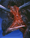
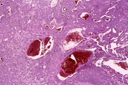
A
B
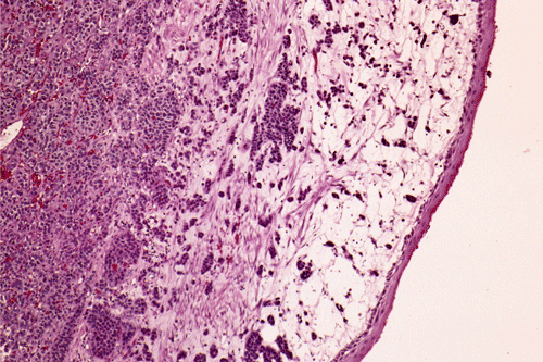
C
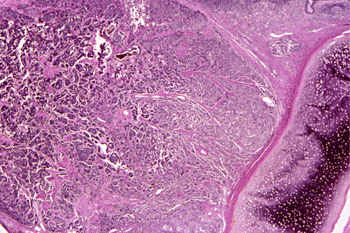
D
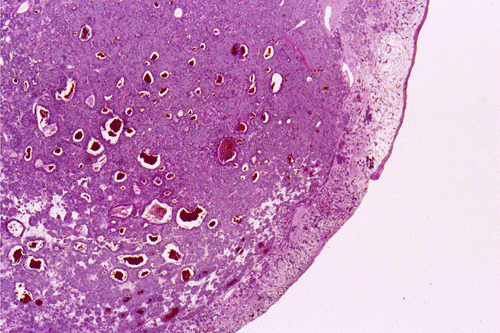
E
F
G
H
I
J
K. Synaptophysin
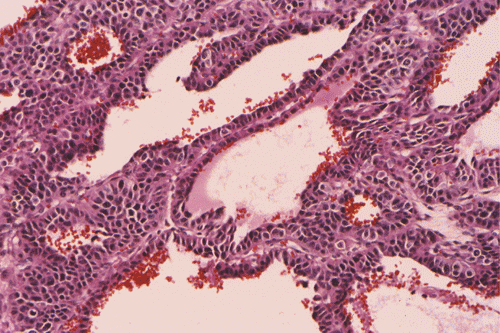
Gross patholgy: The tumor is a pedunculated dark purple nodule that protrude into the lumen of the bronchus (Panel A). The nodular surface is intact and is focally shinny (arrow in Panel A). These type of nodules tend to produce a ball-valve type of effects leading to atelectasis. These areas correspond to mucosal surface with squamous metaplasia (arrow in Panel B).
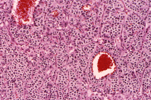
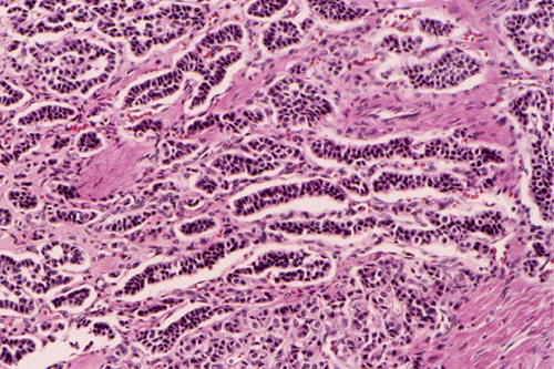
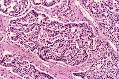
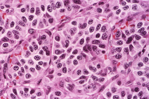
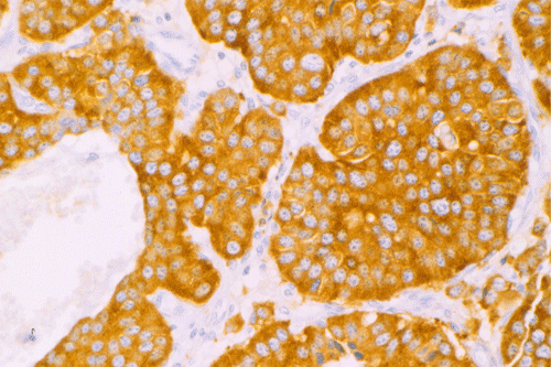
Histopathology: The surface of the tumor nodule is covered partly by epithelium with squamous metaplasia (arrow in Panel B), partly by respiratory type epithelium and partly denuded (Panel C). Even at this magnification, several dilated blood vessels can be seen. The tumor locally invades to the level of cartilage (Panel D). Some dilated blood containing space (Panel E) are present. On higher magnification, some areas arrange in nests (Panel F), cords and trabeculae (Panel G), cribiform (Panel H), and microcysts (Panel I). On high-magnification, the tumor cells appear monotonus, with round nuclei and "salt and pepper" like chromatin, and lacks nucleoli. The cells look epithelioid and have fine, granular, amphophilic cytoplasm. The delicate vascular network around the tumor cell nest is best appreciated at this magnification (Panel J). There are neither significant mitotic activity or necrosis. On immunohistochemistry, the tumor cells are strongly reactive for synaptophysin (Panel K). There are also immunoreactive for cytokeratin.
| DIAGNOSIS: Carcinoid. (well-differentiated neuroendocrine carcinoma) |
Discussion: General Information Pathology Differential diagnosis
General Information
Literally, carcinoid
means something similar to but not the same as carcinoma. This term was first
used by Obendorfer in 1907 to separate a group of tumors in the small intestine
that behave better than conventional carcinomas
1.
Argentaffin granules
were demonstrated in carcinoid cells by Gosset and Masson and suggested that
they arise from Kulchitsky cells
2.
The Kulchitsky cells is finally shown to be a subpopulation of the amine
precursor uptake and decarboxylation (APUD) cells which forms the neuroendocrine
system. Carcinoid tumors are neuroendocrine carcinoma of low-grade malignancy.
These tumors are also known as low-grade neuoendocrine carcinoma to address
their potential malignant behavior.
Because of the diffuse distribution of neuroendocrine cells carcinoid
(well-differentiated neuroendocrine carcinoma) can be found in many organs with
the appendix and small intestine as the most common sites (73%). The
bronchopulmonary system (25.1%) is the most common extragastrointestinal site
3.
pulmonary
carcinoid tumors comprises about 1-2% of all primary bronchopulmonary tumors
4. In
children, the lung is the most commonly involved site followed by the liver
5. Although uncommon in children, carcinoid tumor is
the most common primary endobronchial and pulmonary parenchymal tumor in
children and comprises about one-third of all these tumors. There is no sex
predilection. In contrast to adult bronchopulmonary carcinoids that may remain
asymptomatic, bronchopulmonary carcinoids in children are typically symptomatic.
Clinical manifestations include wheezing
and atelectasis in addition to the characteristic adult triad of cough,
hemoptysis, and pneumonitis. The less common manifestations include weight loss
and hoarseness
6,
7,
8.
Diagnosis may be delayed due to its varied ways of clinical presentations. It is
very important for clinician to aware its existence because early diagnosis and
intervention will improve the outcome of the patient’s management.
Carcinoids occasionally occurs as part of multiple neuroendocrine neoplasm (MEN)
syndrome. Carcinoid may also produce an excess amount of neuropeptides and
amines (hormone-like substances, such as bradykinin, serotonin, histamine, and
prostaglandins, which can cause the
carcinoid
syndrome (flushing,
bluish skin, abdominal cramps, diarrhea, heart damage, and other symptoms). Although
most carcinoids are amenable to resection, they carry a low metastatic
potential. Prognosis is general good with a low rate of metastasis. In adult,
the
5- and 10-year survival rates are 87% and 87%, separately. Prognosis is also
good in pediatric cases hoarseness
7.
Currently,
surgery is considered the only potentially curative therapy in this group of
patient because most carcinoids are resistant to both radiation and
chemotherapy.
Grossly, the majority of these tumors arise as is polypoid, endobronchial
mass that protrude into the bronchial lumen. The main to segmental bronchi are
the most common sites probably because this area has the highest concentration
of neuroendocrine cells in these regions. Entire peripheral tumors in lung
parenchyma can be seen. Grossly, endobronchial tumor has a smooth surface and is
pink to purple because of its rich vascularity. The tumor in the periphery of
the lung presents in a solid and nodular pattern. Carcinoid tumors are
well-circumscribed and contain infrequent areas of hemorrhage. Foci of
calcification or ossification may be present. Necrosis is uncommon and, when
detected, should raise the possibility of a higher grade tumor.
Histological features of typical carcinoid tumors are similar to those of
carcinoid tumors found elsewhere in the body. Architecturally, the tumor cells
form nests, cords, or broad sheets separated by highly vascular septa of
connective tissue, or they may take a glandular or alveolar configuration.
Individual cells are medium-sized polygonal with oval to spherical, rather
uniform, finely granular nuclei and lightly eosinophilic granular to clear
cytoplasm; rosettes and small acinar structure with or without mucin may be
present. Pleomorphism is minimal and mitotic figures are rare. Spindle-shaped
cells are an accepted variant, especially in peripherally located tumors. The
stroma is vascular and scanty, and amyloid deposits with bone formation may be
seen. Small foci of atypical hyperplastic bronchial epithelium may form
tumorlets in adjacent bronchial epithelium location and may represent local
metastatic disease. These lesions may suggest a more aggressive tumor and a
poorer prognosis.
Immunohistochemical findings in carcinoid tumors include the reactivity
to chromogranin A, neuron-specific enolase (NSE), synaptophysin, serotonin,
gastrin, MSH, vasopresin, bombesin, somatostatin.
Neural cell-adhesion molecule (N-CAM) reactivity was detected in all
bronchial carcinoid tumors and S-100 protein is present in the sustentacular
cells situated at the border of cell nest in approximately 40% of cases. They
are also positive for epithelial markers such as cytokeratins. Ultrastructurally,
membrane-bound electron-dense granules 100 nm to 400 nm in diameter (neurosecretory-type
granules) are seen in the cytoplasm beneath the cell membrane, often at the
base. Microvilli and junctional complexes are also present.
Differential diagnosis
Typical carcinoid tumors have their unique macroscopic and microscopic
characteristics and do not usually pose any diagnostic problem.
Immunohistochemical stains and other special stains can be useful in confirming
the neuroendocrine differentiation of suspected carcinoid tumors.
Attention may be attended to the followings:
Atypical carcinoid (moderately differentiated neuroendocrine carcinoma)
refers to carcinoid tumors with anaplastic feature and less favorable prognosis
than typical carcinoid tumor. The 5- and 10 year survival
rates are 56% and 35%. Aside from the typical neuroendocrine morphology
of typical carcinoids, moderately differentiated neuroendocrine carcinoma have
increased mitotic activity (>5 mitoses per 10 high-power fields), pronounced
cytologic atypia and frequent necrosis
9,
10,
11,
12. There is no significant difference in immunohistochemical ultrastructural characteristics between typical and
well-differentiated neuroendocrine carcinoma (atypical carcinoid tumor).
Large cell neuroendocrine carcinoma (LCNEC) and small cell lung carcinoma (SCLC)
are
other two major histological types of neuroendocrine tumors of the lung, along
with typical and atypical carcinoids. They are highly malignant with an
extremely low 5- and 10-year survival rate, 27% and 9% for
LCNEC, 9% and 5% for SCLC, respectively (Shimosato).
LCNEC is a rare tumor that, with the exception of the size of the tumor
cells, is very similar to SCLC in its prognosis and in its treatment.
These two types of tumors often possess certain cellular-chemical
features with neruoendocrine tumors. With the highly anaplastic features of
these tumors, differentiation from typical and atypical carcinoids are not
difficult.
Reference:
Obendorfer
S, FrankfurtZ: Karzinoide tumoren des duenndarms. Pathol 1907 1:426-430.
Gosset
A, Masson P. Tumerus endocrines de l’appendice. Pre med 1914 22:37-40
Modlin
IM, Sandor A.
An analysis of 8305 cases of carcinoid tumors. Cancer, 1997; 813-829.
Shimosato
Y. Pulmonary Neoplasm in Diagnostic Surgical Pathology, edited by
Sternbert SS et al., Lippincott Williams and Wilkins, 3rd ed. pp. 1095.
Broaddus
RR, Herzog CE, Hicks MJ.
Neuroendocrine Tumors (Carcinoid and Neuroendocrine Carcinoma) Presenting at
Extra-appendiceal Sites in Childhood and Adolescence.
Arch
Pathol Lab Med. 2003; 127: 1200-3.
Hancock
BJ, Di Lorenzo M, Youssef S, Yazbeck S, Marcotte JE, Collin PP.
Childhood primary pulmonary neoplasms.
J Pediatr Surg, 1993; 28:1133-1136.
Wang
LT, Wilkins EW Jr, Bode HH.
Bronchial carcinoid tumors in pediatric patients. Chest, 1993; 103:
1426-8.
Arrigoni
MG, Woolner LB, Bernatz PE.
Atypical
carcinoid tumors of the lung. J
Thorac Cardiovasc Surg.
197;64:413-21.
Paladugu
RR, Benfield JR, Pak HY, Ross RK, Teplitz RL.
Bronchopulmonary
Kulchitzky cell carcinomas. A new classification scheme for typical and
atypical carcinoids. Cancer.
1985; 15;55:1303-11.
el-Naggar
AK, Ballance W, Karim FW, Ordonez NG, McLemore D, Giacco GG, Batsakis JG.
Typical
and atypical bronchopulmonary carcinoids. A clinicopathologic and flow
cytometric study. Am
J Clin Pathol.
1991; 95:828-34.
Travis WD, Rush W, Flieder DB, Falk R, Fleming MV, Gal AA, Koss MN. Survival Analysis of 200 Pulmonary Neuroendocrine Tumors With Clarification of Criteria for Atypical Carcinoid and Its Separation From Typical Carcinoid, Am J Surg. Pathol. 1998; 22:934-944.