
Myeloperoxidase
CD68
CD3
CD5
CD20
| A 33 year-old Man with an
Epidural Mass in the Thoracic Spine. January, 2004, Case 401-1. Home Page |
Walter F. Bierbaum, M.D., William F. Kern, M.D. Last update: January 30, 2004.
Department of Pathology, University of Oklahoma Health Sciences Center, Oklahoma City, Oklahoma.
Clinical information:
The patient was a 33 year-old white man who presented with a 6 month history of interscapular and thoracic back pain with substantial worsening in the last three months. He also complained of intermittent numbness in his left leg down to his heel, night sweats and generalized fatigue. Otherwise, he had no other problem with ambulation. There were no bowel or urination problems. Imaging studies revealed a compression fracture of the T7 vertebral body with a left extradural mass at T4-T5 levels. A CT-guided needle biopsy of the lesion yielded non-diagnostic material that contained scant bone marrow tissue with scant atypical cells. A T4-T6 hemilaminectomy with tumor debulking was performed. A bone marrow biopsy was performed. The followings are representative photos taken from the extradural mass and bone marrow biopsy:
|
|
|
|
|
|
|
|
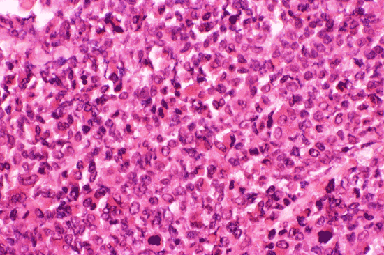
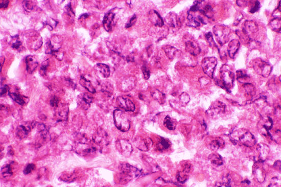
| A. | B. |
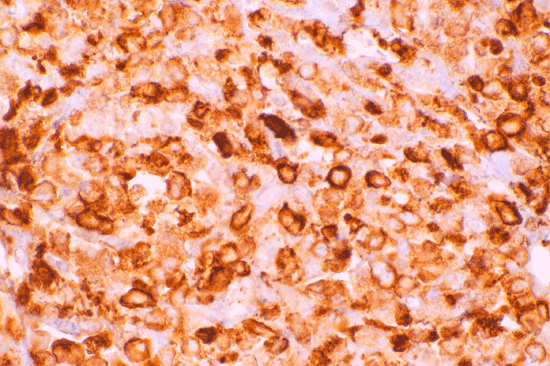
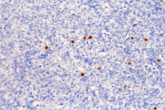
C. Myeloperoxidase |
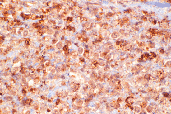
D. CD68 |
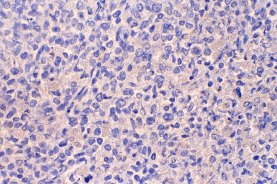
E. CD3 |
F. CD5 |
G. CD20 |
 |
 |
 |
 |
 |
|
|
H. DiffQuick |
I. DiffQuick |
J. DiffQuick |
K. DiffQuick |
L. DiffQuick |
Pathology of the case:
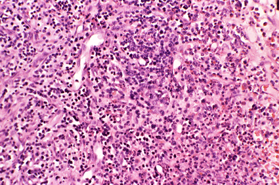
Histopathology: The lesion consisted of solid and cellular areas featuring intermediate to large mononuclear cells admixed with many eosinophils. The mononuclear cells have vesicular chromatin and prominent nucleoli (Panel A and B). Immunohistochemistry disclosed strong reactivity for myeloperoxidase in most of the cells (Panel C) and CD68 (Panel D). Scant cells were immunoreactive for CD3, CD5, and CD20 (Panel E, F, and G).
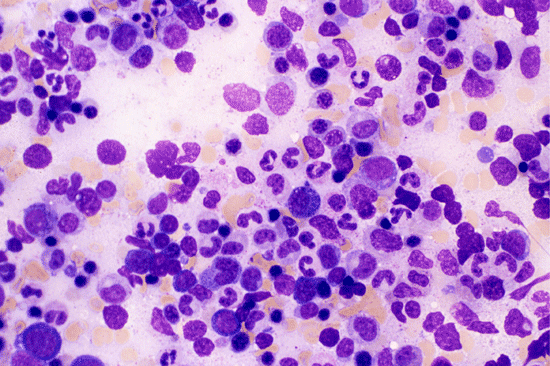
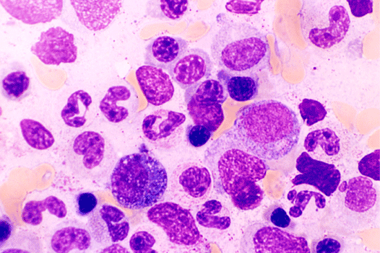
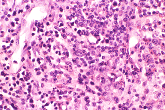
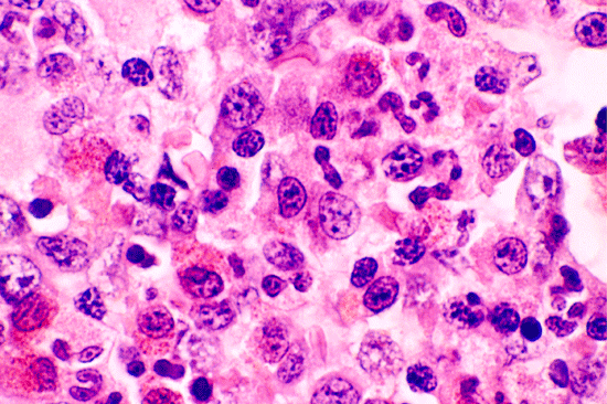
Bone marrow : A heterogeneous population of granulocytic precursors in various stages of maturation (Panels H and I) is revealed in the aspirate. There are no myeloblasts. The bone marrow is hypercellular (approximately 100% cellularity) and contains many large, immature appearing cells and numerous esoinophils (Panels J, K, and L).
Cytogenetics: There is a translocation between chromosomes 8 and 9, involving 9p21. Chromosomal translocation t(8;9) reportedly has an association with chronic myelogenous leukemia.
| DIAGNOSIS: Myeloid sarcoma. |
Discussion: General Information Pathology Differential diagnosis
General Information
Myeloid sarcoma 1, 2 is an extramedullary collection of neoplastic myeloid precursor cells. Different names including chloroma (a term initially used because of the tumor’s transient green appearance which fades on exposure to oxygen), granulocytic sarcoma, and extramedullary myeloid tumor ( a term which describes extramedullary and extrameningeal collections of myelocytic precursors) have been used to describe this condition 3, 4. Although a small number of myeloid sarcoma occur de novo (i.e. with the absence of acute myeloid leukemia, myelodysplastic syndrome, or other myeloproliferative disorder in a bone marrow biopsy within 30 days of the diagnosis of myeloid sarcoma), most of them occur with concurrent bone marrow involvement by a myelodysplastic syndrome or a myeloproliferative disorder, most commonly acute myeloid leukemia. The lesion also provides evidence of relapsed acute myeloid leukemia with or without concurrent bone marrow involvement. Occasionally, there may be a prolonged interval (months to years) between the occurrence of myeloid sarcoma and acute myeloid leukemia.
Myeloid sarcomas may arise almost anywhere in the body. Subperiosteal tissue of the skull and paranasal sinuses, sternum, ribs, vertebrae and pelvis are the most common sites. The skin and lymph nodes are also affected. Children up to the age of 16 with acute myeloid leukemia have a proportionately higher incidence of orbital myeloid sarcoma compared to adults 5. Children with acute myelomonocytic leukemia also seem to have a high incidence of orbital involvement with exophthalmos, chemosis and orbital masses as symptoms 6.
The overall incidence of myeloid sarcoma is difficult to determine secondary to lack of uniform biopsy standards and uncertain correlation of abnormal physical findings. Retrospective studies report an incidence between 3% and 20% (in a study which described only meningeal leukemias). The incidence reports varied based on the ages of the patients studied and sites excluded from the analysis (most commonly skin or meninges) 6. Myeloid sarcomas, in general, occur in a relatively larger percentage of children with acute myeloid leukemia, particularly infants (< 2 years old). Patients with myeloblastic and monoblastic (French-American-British (FAB) M4) and monoblastic (FAB M5) leukemia tended to have a higher incidence of myeloid sarcoma, particularly leukemia cutis and gingival involvement, than patients in other FAB classifications 6, 7, 8, 9.
There are two major types of myeloid sarcoma- the granulocytic sarcoma and monoblastic sarcoma. The granulocytic sarcoma is the most commone one and is composed of myeloblasts, neutrophils and precursors of neutrophils. The degree of maturation ranges from blastic, immature to differentiated. The blastic type is composed primarily of myeloblasts while the differentiated type is composed of more mature neutrophils and promyelocytes. The immature type is composed of a mixture of myeloblasts and promyelocytes. The monoblastic sarcoma is predominantly of monoblasts. Tumors with tri-lineage hematopoiesis, a predominance of erythroid precursors, or a predominance of megakaryocytes may occur in conjunction with transformation of chronic myeloprotive disorders 2. Imprints are very helpful tool for diagnosis particularly for intraoperative consultations.
Immunohistochemical and/or histochemical demonstration for the presence of myeloperoxidase, lysozyme and chloroacetate esterase are critical for identification of their myeloid origin. An immunohistochemical profile is also helful in subtyping these tumors 10, 11. Genetic translocations that are observed in acute myeloid leukemia may also be demonstrated in myeloid sarcomas. In our case, there is a translocation between chromosomes 8 and 9, involving 9p21. T(8;9) reportedly has an association with chronic myelogenous leukemia 12. Association of myeloid sarcoma with acute myeloid leukemia with maturation and t(8;21)(q22;q22) and acute myelomonoblastic leukemia with abnormal eosinophils with inv (16)(p13p22) and (p16;16)(p13;q22) may occur 2.
Differential diagnosis
The differential diagnoses for myeloid sarcoma includes Langerhans cell histiocytosis (particularly in those cases which have a prominent eosinophilic infiltrate), lymphomas (particularly diffuse large B cell lymphoma), peripheral neuroectodermal tumor/ Ewing’s sarcoma.
Langerhans cell histiocytosis may be considered in the differential diagnosis, particularly when the tumor has an eosinophilic infiltrate. The degree of pleomorphism in Langerhans cell histiocytosis is usually less prominent than myeloid sarcoma and their characteristic oval nuclei with longitudinal grooves, the so-called “coffee bean” appearance, allows morphologic separation between the two entities. The Langerhans cells are also positive for S-100 protein and CD1a.
Various types of lymphoma may be extremely difficult to differentiate from myeloid sarcoma. Demonstration of myeloid phenotype is critical for the distinction. In soft tissue sites traditionally known to harbor lymphoma (e.g. testes or lymph nodes) concurrent immunohistochemistry or flow cytometry frequently is helpful in making the diagnosis of lymphoma. A battery of immunohistochemical stains, including lymphocyte common antigen (CD45) or a B-cell marker such as CD20 could circumvent the potentially disastrous mislabeling of a myeloid sarcoma.
Peripheral neuroectodermal tumor/ Ewing’s sarcoma is a one of the “small blue cell tumors” and is the second most frequent solid bone tumors among children and adolescents. Clininal and radiographic correlation is extremely important for the correct diagnosis of this lesion. The tumor frequently consists of monotonous round cells with hyperchromic nuclei and scant cytoplasm. In more than 85% of cases, the fusion gene EWS/FLI-1 from t(11;22) may be identified from cytogenetic analysis or polymerase chain reaction. The tumor frequently expresses CD99.
Reference:
Byrd JC, Edenfield WJ, Shields DJ, Dawson NA. Extramedullary myeloid cell tumors in acute nonlymphocytic leukemia: a clinical review. J Clin Oncol. 1995 13(7):1800-16.
Brunning RD, Matutes E, Flandrin G et al. Acute myeloid leukaemia not otherwise categroised. In Jaffe ES, Harris NL, Stein H, Vardiman JW, WHO Classification of Tumors: Tumors of Hematopoietic and Lymphoid Tissues. IARC Press. Lyon. 2001: 104-105.
Schultz J et al. The Chemistry of experimental chroroma: I. Prophyrins and peroxidases. Cancer Res 1954; 14:157-162.
Rappaport H: Tumors of the hematopoetic system, in Atlas of Tumor Pathology, Section III, Fascicle 8. Washington DC, Armed Forces Institute of Pathology, 1967: 241-247.
Shome DK, Gupta NK, Prajapati NC, Raju GM, Choudhury P, Dubey AP. Orbital granulocytic sarcomas (myeloid sarcomas) in acute nonlymphocytic leukemia. Cancer. 1992 70(9):2298-301.
Cavdar AO, Babacan E, Gozdasoglu S, Kilicturgay K, Arcasoy A, Cin S, Ertem U, Erten J. High risk subgroup of acute myelomonocytic leukemia (AMML) with orbito-ocular granulocytic sarcoma (OOGS) in Turkish children. Retrospective analysis of clinical, hematological, ultrastructural and therapeutical findings of thirty-three OOGS. Acta Haematol. 1989 81:80-5.
Tobelem G, Jacquillat C, Chastang C, Auclerc MF, Lechevallier T, Weil M, Daniel MT, Flandrin G, Harrousseau JL, Schaison G, Boiron M, Bernard J. Acute monoblastic leukemia: a clinical and biologic study of 74 cases. Blood. 1980 55:71-6.
Shaikh BS, Frantz E, Lookingbill DP. Histologically proven leukemia cutis carries a poor prognosis in acute nonlymphocytic leukemia. Cutis. 1987 39:57-60.
Baer MR, Barcos M, Farrell H, Raza A, Preisler HD. Acute myelogenous leukemia with leukemia cutis. Eighteen cases seen between 1969 and 1986. Cancer. 1989 63:2192-200.
Audouin J, Comperat E, Le Tourneau A, Camilleri-Broet S, Adida C, Molina T, Diebold J. Myeloid sarcoma: clinical and morphologic criteria useful for diagnosis. Int J Surg Pathol. 2003 11:271-82.
Davey FR, Olson S, Kurec AS, Eastman-Abaya R, Gottlieb AJ, Mason DY. The immunophenotyping of extramedullary myeloid cell tumors in paraffin-embedded tissue sections. Am J Surg Pathol. 1988 12:699-707.
Borker A, Yu L, Ode D. Blast crisis of chronic myeloid leukemia: diagnosis prompted by T(8;9). J Pediatr Hematol Oncol. 2002 24:670-1.