| A 13 month-old Girl with a
Mass in Temporal Bone. February, 2004, Case 402-2. Home Page |
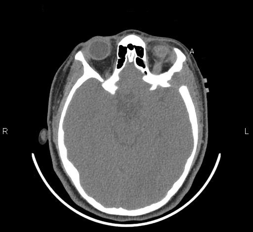
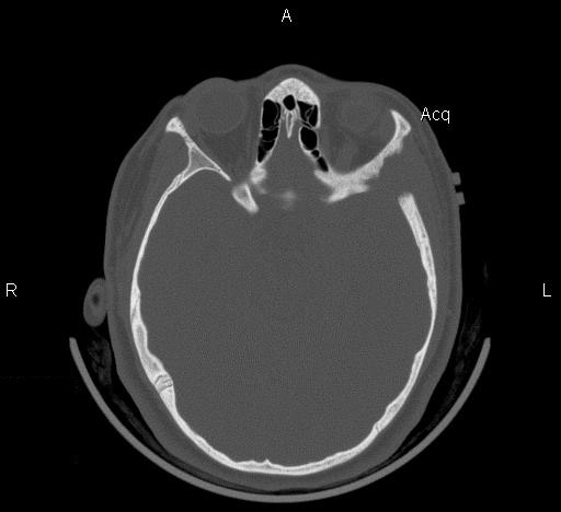
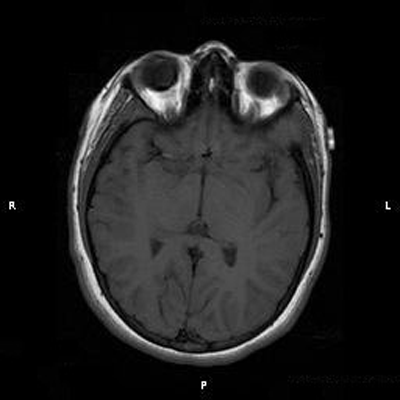
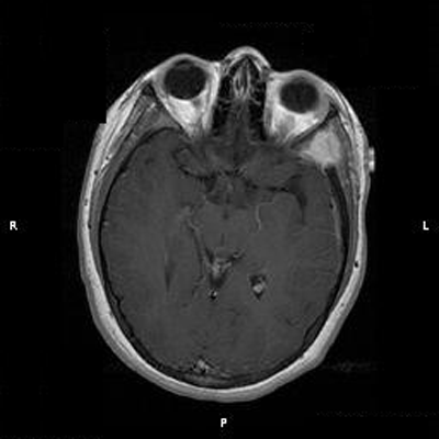
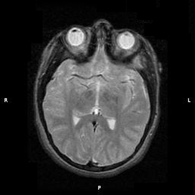
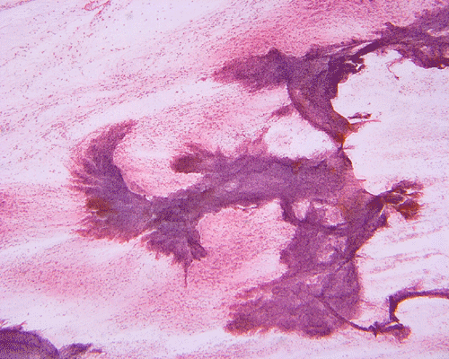
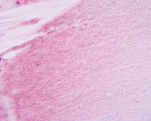
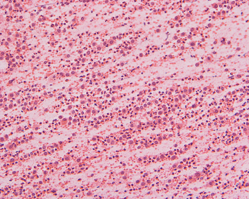
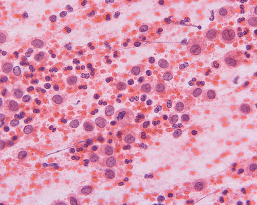
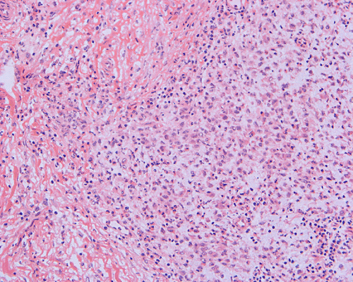
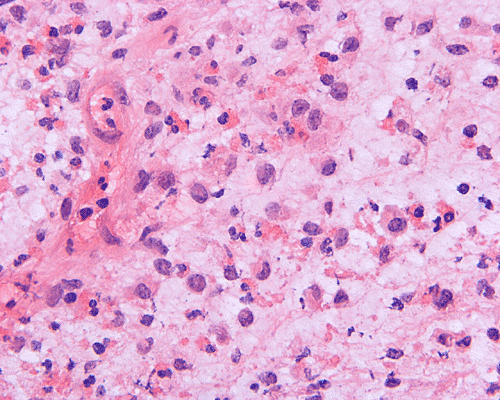
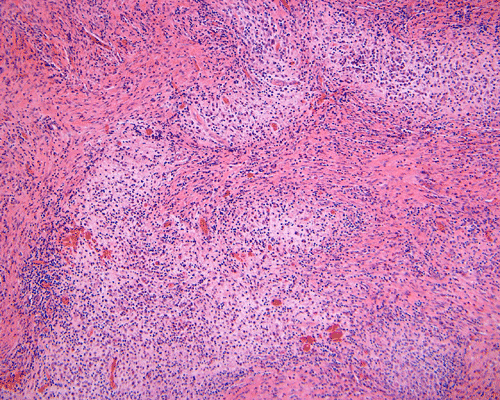
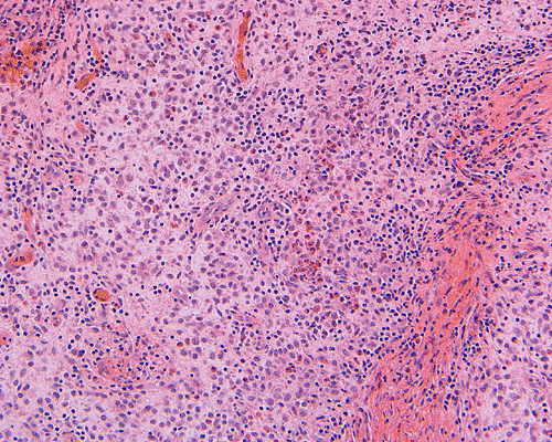
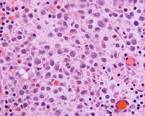
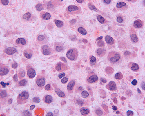
Clinical information: The patient was a 13 year-old girl who presented with worsening headache and some difficulties with memory, concentration and attention. MRI studies disclosed a 2.1 x 1.9 x 1.8 cm enhancing mass in the left temporal bone and sphenoid wing. The mass extended intracranially and abuts the left temporal bone accompanied by dural enhancement at that location. There is also extension through the bone into the submuscular temporal region. The following photos are taken from representative regions of the lesion. Panel 1 and 2 are CT scans at soft tissue and bone density respectively. Panel 3 and 4 are T1 weighed images without and with contrast respectively. Pandl 5 is proton density image. Panel A to D are cytologic (squash) prepartion for intra-operative consultation. Panel E and F are frozen sections for intraoperative consultation. Panel G to I are paraffin embedded sections.
|
|
|
|
|
|
|
| 1. | 2. | 3. | 4. | 5. |
 |
 |
 |
 |
 |
|
|
A. Squash |
B. Squash |
C. Squash |
D. Squash |
E. Frozen |
|
 |
 |
 |
 |
 |
|
|
F. Frozen |
G. | H. | I. | J. |