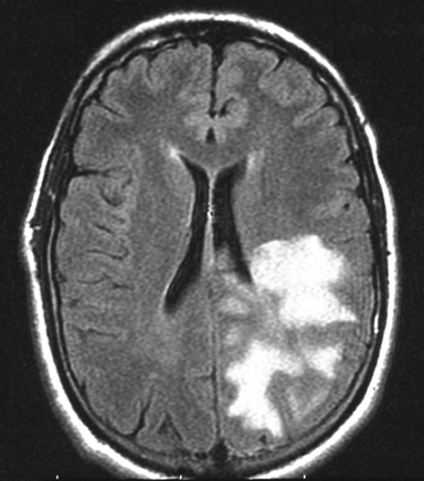
FLAIR
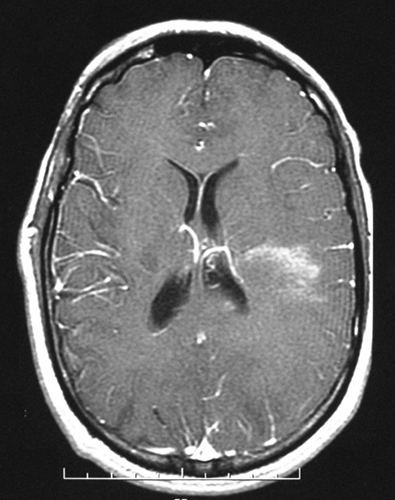
T1 + Contrast
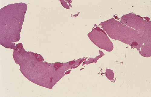
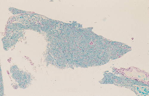
LFB/PAS
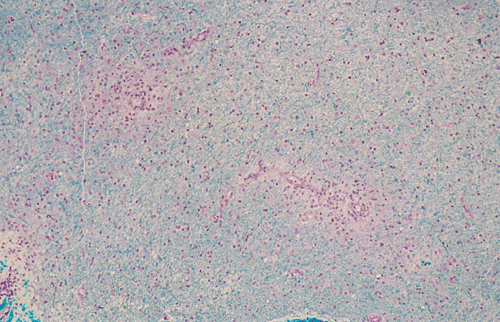
LFB/PAS
LFB/PAS
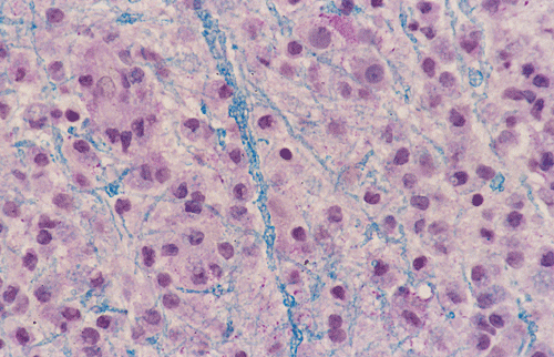
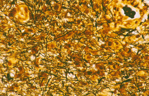
Biel
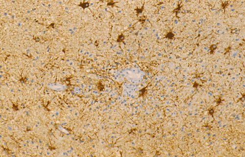
GFAP
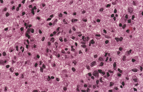
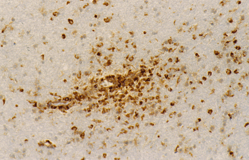
CD68
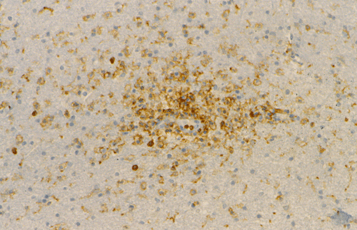
LCA
| A 27 year-old Man with
Headache, Visual Field Loss and Right Side Weakness. July, 2004, Case 407-2. Home Page |
Amir Ilyas, M.D.1, Kalliopi Petropoulou, M.D. 2, Zahid F. Cheema, M.D.1, Kar-Ming Fung, M.D., Ph.D.3 Last update: February 28, 2005..
1 Department of Neurology, 2 Department of Radiology, 3 Department of Pathology, University of Oklahoma Health Science Center, Oklahoma City, Oklahoma
Clinical information: The patient was a 28 years old ambidextrous, previously healthy Caucasian man. He was admitted to the hospital because of new onset headache and right sided hemiparesis. The patient experienced a flu like symptoms associated with high fever and chills for 3 weeks a few months before admission. His symptoms resolved and he remained well for 2 months. Three weeks before admission, he started to have severe occipital headache with a pulsating nature and worsened with coughing. The headache was not initially associated with nausea, vomiting, photophobia, phonophobia or visual symptoms. Later, he developed difficulty in visualizing objects in his right visual field followed by weakness in his right upper extremities, lower extremities, and right face dropping. He then experienced slurred speech, difficulty in writing and word finding. He was admitted to the hospital.
On physical examination, his higher mental functions were all intact. There was significant visual field cut in the form of almost complete right hemianopsia. There was right facial drop consistent with lesions in the upper motor neurons. The left upper extremities had 4/5 strength and the lower extremities had a 5/5 strength. The right upper extremities had a 3/5 strength and the lower extremities had a 3/5 strength. His planter reflex on the right side was upgoing with brsik reflexes and was normal and down going on the left. Laboratory study on his cerebral spinal fluid was within normal limits and no oligoclonal bands were identified. Laboratory study on ANA, lyme disease, Toxoplasma, HIV, JC virus, herpes simplex virus, varicella zoster virus, West Nile virus, and St. Louis encephalopathy were negative.
The patient became more confused and incoherent. A CT scan showed a mass lesion with edema on the left side of the brain that was suggestive of a tumor. MRI showed a large infiltrating lesion involving the left parietal lobe, occipital lobe, a portion of the parietal lobe, and part of the basal ganglia. The lesion was mildly enhancing after gadolinium injection. A stereotactic biopsy was performed. Representative MRI images and histologic images are illustrated below.
|
|
|
|
|
|
|
|
A. FLAIR |
B. T1 + Contrast |
C. | D. | E. |
F. LFB/PAS |
|
|
|
|
|
|
|
|
G. LFB/PAS |
H. LFB/PAS |
I. Biel |
J. GFAP |
K. CD68 |
L. LCA |
MR Imaging of the case:
The FLAIR sequences (Panel A) demonstrate an extensive abnormality with increased intensity signal and irregular borders that involves the left occipital, parietal and dorsal frontal lobes. Although the lesion is quite extensive the mass effect is minimal. On the post contrast T1-weighted sequences (Panel B) there is enhancement only in the anterior border of the lesion. The limited mass effect along with the “crescent” rim enhancement are suggestive of demyelinating process, likely tumefactive multiple sclerosis.
Pathology of the case:
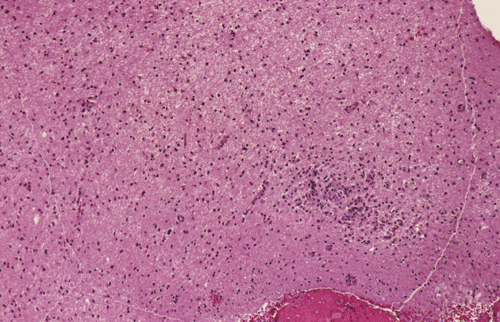
Streotactic biopsy yielded several cores of tissue. On scanning magnification, there are multiple small lymphocytic collections (Þ in Panel C) in the white matter. On higher magnification, these areas contain mostly lymphocytes without atypia. These collections appear to be angiocentric and are often admixed with some foamy macrophages (Panel D and E). On LFB/PAS stain, multiple small, irregular areas of angiocentric pale staining are present (Panel F and G). On high magnification, these areas are featured by infiltration by foamy macrophages and lymphocytes and myelin loss (Panel H). On Bielschowsky stain, the axons in these pale areas are largely preserved. Immunohistochemistry on GFAP demonstrates multiple stellate reactive astrocytes (Panel J). The macrophages and lymphocytes are also well demonstrated by immunohistochemistry for CD68 and LCA respectively (Panel K and L).
Comment:
From the imaging point of view, the involvement is extensive and without mass formation. The lesion also touches the ventricles. These features are compatible but not diagnostic of an inflammatory and demyelinating process. The salient pathologic differential diagnoses of this case is demyelinating disease versus infections versus lymphoma. Obviously, there is a lack of angiocentric arrangement of atypical lymphocytes that is typical for primary lymphoma of the central nervous system (CNS). The clinical findings do not suggest secondary involvement of the brain by a lymphoma or leukemia. If an infectious agent is involved in this process, it would most probably be a virus. Occasionally, progressive multifocal leukoencephaly (PML) in patients with HIV infection may manifest as an initial presentation in a "previously healthy" patient. Some of these cases may present with a single lesion and therefore clinically suggesting a neoplastic process. It is, therefore, important to look for viral inclusions and atypical cells in cases with the current histopathologic findings. PML is often not purely demyelinating but admixed with some necrosis. Laboratory studies of the cerebral spinal fluid (CSC) and blood are also helpful in identifying the viral agent. Electrophoresis for oligoclonal band in the CSF are often helpful in diagnosing demyelinating diseases.
Aftermath:
This patient was treated by steroid and completely recovered. There was no relapse in the two year follow up period.
| DIAGNOSIS: Inflammatory demyelinating pseudotumor |
Discussion: General Information Pathology ADEM vs. MS
General Information
Inflammatory demyelinating diseases have a spectrum of manifestations in the central nervous system (CNS). On one end of the spectrum, they would occur as an acute, single episode (monophasic), global process with acute disseminated encephalomyelitis (ADEM) as the prototype. While the classic type occur as a acute disseminated perivenous encephalomyelitis, the hyperacute type occurs as acute hemorrhagic encephalitis. On the other end of the spectrum, it would occur as a chronic recurrent, relapsing process with the Charcot or classic type of multiple sclerosis (MS) as the prototype. It has been well documented that multiple sclerosis may clinically mimic neoplasms 1, 2, 3. Kepes described a series of large, focal tumor-like demyelinating lesions of the brain. In his series, many of the cases were unifocal, single episode (monophasic) lesions. These features suggested that these lesions are intermediate lesions between multiple sclerosis and acute disseminated encephalomyelitis 4. In his study of 31 patients aged between 10 years to 77 years, 24 patients had unifocal and 7 patients had multifocal lesions. No additional lesions developed in 28 patients after a 12 year period of follow up. There were three patients who develop additional lesions consistent with MS in follow up between 9 months to 12 years. Inflammatory demyelinating pseudotumor seems to be an appropriate name for this type of lesions. Similar cases have been reported subsequently 5, 6. It is very important not to mistaken an inflammatory demyelinating process as tumor as the consequences of radiation therapy on patients with demyelinating disease are catastrophic 7.
The histopathology of inflammatory demyelinating pseudotumors is very similar to that of acute disseminated perivsnous encephalomyelitis or the classic type of ADEM. In essence, there is perivenous chronic inflammatory cell infiltration accompanied by infiltrating, foamy macrophages with an angiocentric distribution. Loss of myelin and relative preservation of axon fibers must be demonstrated before a diagnosis of demyelinating process is made. The loss of myelin can be well demonstrated by special stains for myelin with the Luxol fast blue based stains as one of the most commonly used stains. Relative preservation of axons can be sell demonstrated by silver stains such as Bodian stain, Bielschowsky stain, and also immunohistochemistry for moderate and high molecular weight neurofilament proteins. The pathologic highlights on separating inflammatory demyelinating lesions from mimicking lesions have been well described by Zagzag et al. 8. Other inflammatory lesions, particularly viral infectons, must also be ruled out. Keen observations particularly for nuclear inclusion bodies, immunohistochemistry for specific viral agents, and laboratory studies of the cerebral spinal fluid (CSF) and blood are often helpful. Identification of oligoclonal bands on electrophoresis of CSF is a strong suggesting of a demyelinating process.
Acute disseminated encephalomyelitis (ADEM) versus multiple sclerosis (MS)
ADEM is typically a monophasic inflammatory demyelinating disease of the CNS involving brain and spinal cord which often follows either infections or after vaccinations. Some of the most common infectious agents that would trigger ADEM include measles rubella, mumps, varicella zoster, Mycoplasma, and vaccinations against measles, ,mumps, rubella and rabies. ADEM is typically seen in children and young adults and it accounts for approximately 20% of acute cases of encephalitis in children. Male patients are more affected then female patients. An autoimmune type of mechanism has been suggested.
Clinical manifestations including fever, malaise headache, nausea and vomiting often preceded the neurological symptoms which would include parasthesias, pain, motor weakness, aphasia, incoordination, dysarthria ataxia. MRI shows multiple patchy areas of increase signal intensity on Conventional T2 weighted and proton density weighted images and FLAIR images can be seen in subcortical white matter, but sometime also involving the spinal cord or brain stem. With T1-weighted image the active lesion can be enhancing and imply breakdown of the blood brain barrier. Sometime grey matter involvement, particularly the basal ganglia and thalamus, can occur. Pathologically, the lesions are of in the same phase. Treatment includes IV steroids, IV-Ig infusion as well as plasmapharesis all showed promising results. Recovery begins within days but mostly occur in weeks to months. Mortality for the classic type of ADEM varies between 10-25% depending on the severity and abruptness of onset and clinical syndrome. Complete recovery occurs in 50% -70%. Measles virus associated ADEM carries a worse prognosis.
MS, on the other hand, are not heralded by preceding infection or vaccination. In about 90% of the cases, the patient follows a chronic, remitting-relapsing course. In about 10% of the cases, the patient follows a continuous downhill course with no remission. Female patients are more affected. Although MS tend to occur in young adults, they are quite uncommon in children. In childhood cases, they are ten times more frequent in girls then in boys. The clinical severity may not be proportional to the volume of affected tissue. Rather, small lesions located at a strategic location such as the pyramid or internal capsule will cause impressive clinical manifestations. Involvement of the thalamus and basal ganglia, although common for ADEM, are uncommon in MS. MRI is a very sensitive but not specific test for the diagnosis of MS. Enhancement can be observed in MS and this particular feature, when it occur in a single lesion, often raise the clinical suspicion of neoplasm 9. Pathologically, MS is featured by multiple lesions are in different stages of development. While the early plaques are featured by active myelin destruction, chronic inflammation and infiltration my macrophages, the old or “burnt-out” plaques are featured by loss of myelin and gliosis. Recovery is variable and typically incomplete in severe cases.
Reference:
Kumar K, Toth C, Jay V. Focal plaque of demyelination mimicking cerebral tumor in a pediatric patient. Pediatr Neurosurg. 1998 29:60-3.
Hunter SB, Ballinger WE Jr, Rubin JJ. Multiple sclerosis mimicking primary brain tumor. Arch Pathol Lab Med. 1987 111:464-8.
Friedman DI. Multiple sclerosis simulating a mass lesion. J Neuroophthalmol. 2000 20:147-53.
Kepes JJ. Large focal tumor-like demyelinating lesions of the brain: intermediate entity between multiple sclerosis and acute disseminated encephalomyelitis? A study of 31 patients. Ann Neurol. 1993 3:18-27.
Heyman D, Delhaye M, Fournier D, Mercier P, Rousselet MC, Menei P. Pseudotumoral demyelination: a diagnosis pitfall (report of three cases). J Neurooncol. 2001 54:71-6.
Erana-Rojas IE, Barboza-Quintana A, Ayala AG, Fuller GN. Demyelinating pseudotumor. Ann Diagn Pathol. 2002 6:265-71.
Peterson K, Rosenblum MK, Powers JM, Alvord E, Walker RW, Posner JB. Effect of brain irradiation on demyelinating lesions. Neurology. 1993 43:2105-12.
Zagzag D, Miller DC, Kleinman GM, Abati A, Donnenfeld H, Budzilovich GN. Demyelinating disease versus tumor in surgical neuropathology. Clues to a correct pathological diagnosis. Am J Surg Pathol. 1993 17:537-45.
He J, Grossman RI, Ge Y, Mannon LJ. Enhancing patterns in multiple sclerosis: evolution and persistence. AJNR Am J Neuroradiol. 2001 22:664-9.