T1
T1+Contrast
T1+Contrast
T2
T1+Contrast
Squash
Squash
Frozen
Frozen
Frozen
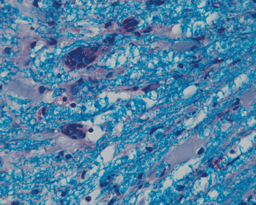
NFP
LFB-PAS
| A 10 year-old Boy with
Asthma and Wearness in Lower Extremities. January, 2005, Case 501-1. Home Page |
Waseem Ahmad, M.D. 1, Kalliopi Petropoulou, M.D.2, Kar-Ming Fung, M.D., Ph.D.3 Last update: August 1, 2005.
1 Department of Neurology, 2 Department of Radiology, and 3 Department of Pathology, University of Oklahoma Health Sciences Center, Oklahoma City, Oklahoma.
Clinical information: The patient was a 9 year-old African-American boy. He developed left lower extremity progressive weakness 4 months ago (in May). As per his father, the patient started "dragging" his left leg in May. He was first evaluated by the orthropedic service and was later referred to the neurosurgery service of this institution. The patient had asthma and his treatment had been intermittent. There was no history of seizures or other neurologic disease. The family history was unremarkable. He was born in the United States never traveled beyond the state border of Oklahoma.
On physical examination, the patient was awake, alert, and well oriented to time, place , person, and situation. There was no memory or f language impairment or mental retardation. Strength in both upper extremities were 5/5 bilaterally. There was also a 5/5 strength in extension and flexion on the right lower extremity. His strength for left hip flexion, extension, abduction and adduction was 5/5. In his left knee, the strength for extension was 5/5 and flexion was 4/5. Strength on left ankle dorsiflexion was 4/5, planter flexion was 2/5, inversion and eversion were 5/5. His peripheral sensation was intact. The reflexes were increased to 3/4 in lower extremities with ankle clonus on the left. There were also bilateral up going toes. There is no impairment on sphincter functions. There was not history or skin rash or insect bite. The remaining of the physical examination was unremarkable. An MRI followed by a biopsy were performed.
Laboratory medicine: The peripheral blood complete and differential count was within normal limits. In particular, no eosinphilia was demonstrated in peripheral blood. There was no evidence of eosinophilia in peripheral blood. A spinal tap was performed before the surgery and reviewed substantial increase in eosinophils. One g-band was also detected in the cerebral spinal fluid (CSF) which suggest an inflammatory process. There was also increase in the level of IgE in the CSF. These information were withheld to increase the challenge level of this case. The results of laboratory testing were as follows:
| Tests on CSF | Result | Reference |
| Color | Colorless | Colorless |
| Appearance | Clear | Clear |
| WBC | 3 | 0-5 /mm3 |
| RBC | 7 | 0 /mm3 |
| RBC appearance | Fresh and crenated | |
| Polymorphonuclear leukocyte | 1 | 0-6% |
| Lymphocyte | 71 | 40-80% |
| Monocyte | 4 |
15-45%
|
| Eosinophil | 24 | 0-2% |
| IgG | 3.5 | 0.0-8.6 mg/dL |
| Glucose | 56 | 60-80 mg/dL |
| Protein | 26 | 15-45 mg/dL |
| Myelin basic protein | 1.8 | 0.0-2.2 ng/nL |
| Oligoclonal band |
One g-band
detected.*
|
*This band is not observed in serum and is, therefore, is of primary CSF origin. Although it is not thought to represent supportive evidence for multiple sclerosis, it may reflect a CNS inflammatory process.
|
Immunology |
Result | Reference |
| IgE | 138 | 0-87IU/ml |
| IgG | 1030 | 691-1539 mg/dl |
| IgA | 179 | 74-214 mg/dl |
| IgM | 69 | 45-153 gm/dl |
| HIV profile | Negative | |
| ANA | 1:40 Nuclear | |
| Mycoplasma IgM | Negative | |
| Toxocara antibody | Negative | |
| Lyme disease panel | Negative |
Representative images from the MR studies and specimen are illustrated below:
|
|
|
|
|
|
|
|
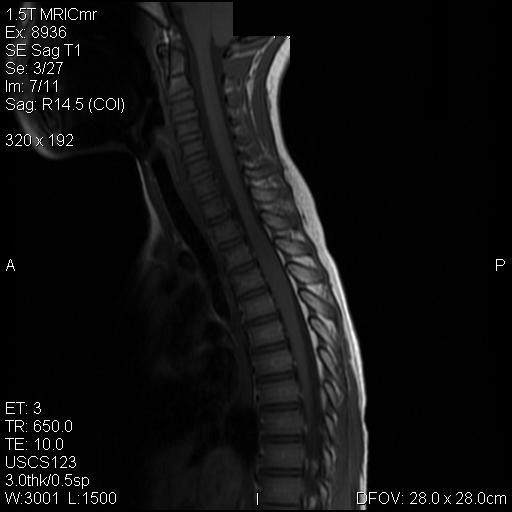
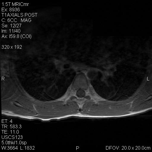
A. T1 |
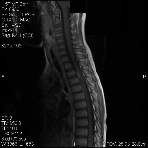
B. T1+Contrast |
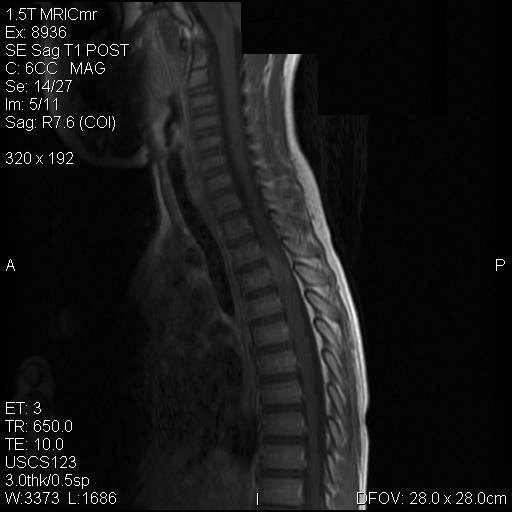
C. T1+Contrast |
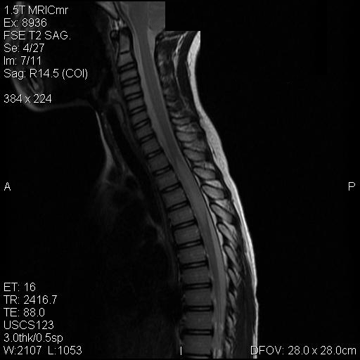
D. T2 |
E. T1+Contrast |
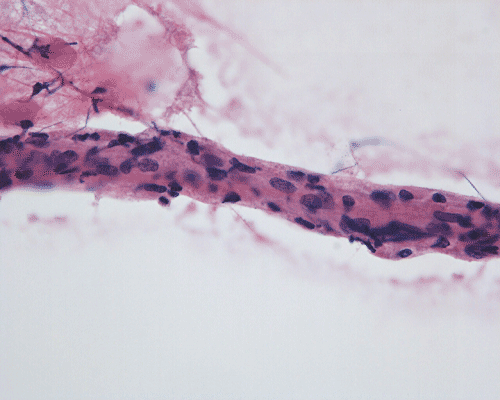
F. Squash |
|
|
|
|
|
|
|
|
G. Squash |
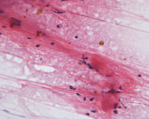
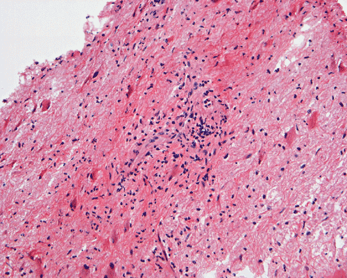
H. Frozen |
I. Frozen |
J. Frozen |
K. | L. |
|
|
|
|
|
|
|
| M. | N. | O. |
P. NFP |
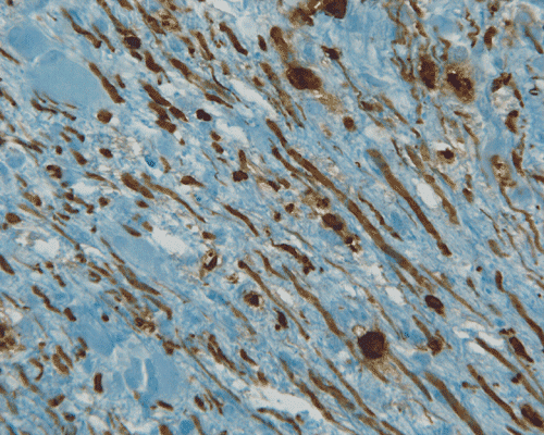
Q. LFB-PAS |
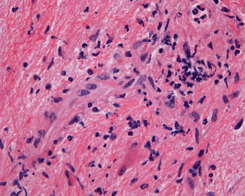
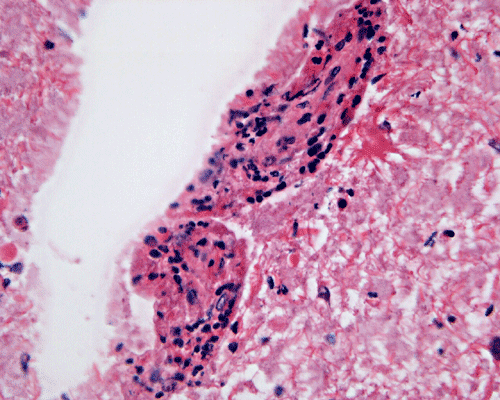
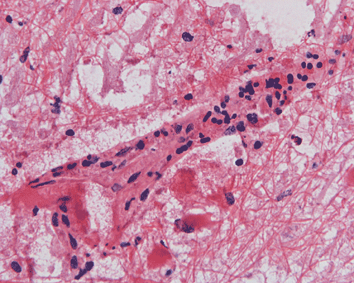
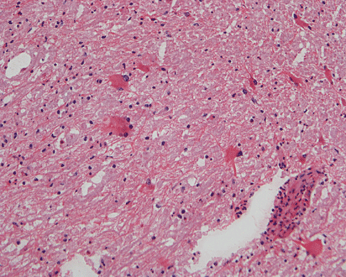
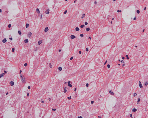
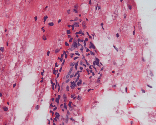
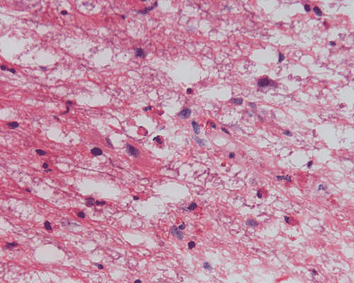
Panel A to E are MR images. Panel F and G are taken from intraoperative cytologic preparation. Panel H to M are taken from frozen section. Panel N and O are from paraffin section. These sections are stained by hematoxylin-eosin stain. Panel P is immunohistochemistry for neurofilament proteins. Panel Q is stained by Luxol fast blue-Periodic acid Schiff (LFB-PAS) stain.
MR Imaging:
The segment of spinal cord at T3-T4 level is expanded and is accompanied by edema that runs from T2-T6. The expansion and edema is roughly symmetrical along the long axis of the cord. The expanded levels are isointense to the white matter of the spinal cord on T1-weighed images (Panel A). The edema is best demonstrated on T-weighed images (Panel B, C, and E). Patchy and poorly defined enhancement is demonstrated in the expanded areas (Panel D). The remaining levels of the spinal cord and the brain are free of abnormal changes.
Pathology:
| DIAGNOSIS: Eosinophilic myelitis. |
Discussion: Acute Myelitis, asthma, & Hopkins sybdrome Pathology Differential diagnosis
Acute transvers myelitis (ATM)
Several cases of “acute myelitis” were described in the nineteenth century, and pathologic analysis revealed that some were due to vascular lesions and others to acute inflammation. In 1922 and 1923, physicians in England and Holland became aware of a rare complication of smallpox vaccination: inflammation of the spinal cord and brain. Given the term post-vaccinal encephalomyelitis, over 200 cases were reported in those two years alone. Pathologic analyses of fatal cases revealed inflammatory cells and demyelination.” In 1928, it was first postulated that many cases of acute myelitis are “post-infectious rather than infectious in cause” since the signs and symptoms of myelitis appear after the fever and rash had resolved. It was proposed, therefore, that the myelitis was an “allergic” response to a virus rather than the virus itself that caused the spinal cord damage. It was in 1948 that the term “acute transverse myelitis” (ATM) was utilized in reporting a case of fulminant inflammatory myelopathy complicating pneumonia.
ATM refers to an acute inflammation of the spinal cord that is restricted to a few segments but extends horizontally across most of the spinal cord of the involved segment. Both sensory and motor functions are affected and the thoracic cord is most commonly involved.. The term "transverse" is not very precise from the clinical point of view as the spinal manifestations are often asymmetrical. In many circumstances, it is a manifestation of multiple sclerosis. However, ATM can occur in many different situations including autoimmune diseases such as systemic lupus erythematosis, autoimmune vasculitis, Lyme disease, syphilis, radiation and schistosomiasis. ATM can also be resulted from direct viral infection and post-infectionous reaction. Myelitis, sometimes in the form of transverse myelitis, and encephalomyelitis remain well documented but rare complications to a variety of vaccinations 1, 2, 3, 4. Clinically, there is rapidly progressive (in terms of hours or days) paraparesis, sphicteric dysfunction, and bilateral Babinski sign. The acute onset is marked by flaccid paralysis of the lower extremities with acute urinary retention. The sensory level is usually a good guide of the level being involved.
Comment on this case: In our case under discussion, there is myelitis involving a relative short segment of the spinal cord. There is lost of some motor functions but sensory functions are preserved. The course evolves over a couple of months. Although it has somve features of transverse myelitis, it is not acute and should not be regarded as ATM.
Myelitis, asthma, and Hopkins syndrome
ATM is a group of disorders characterized by focal inflammation of the spinal cord and resultant neural injury. It is rare and its incidence is about 1.34 per million population 5. A history of infection was present in 37% of the patients in one study and are more common among younger patients 5. ATM may be an isolated entity or may occur in the context of multifocal or even multisystemic disease. It is clear that the pathologic substrate-injury and dysfunction of neural cells within the spinal cord- may be caused by a variety of immunologic mechanisms. For example, in ATM associated with systemic disease (i.e. systemic lupus erythematosus or sarcoidosis), a vasculitic or granulomatous process can often be identified. In pathologic processes that involves substantial damage of the blood vessels, thrombosis of the damaged vessels may occur and add ischemic damage to the inflammatory pathologic process. There is intraparenchymal and/or perivascular cellular influx into the spinal cord resulting in breakdown of the blood-brain barrier and variable demyelination and neuronal injury.
Eosinophilic myelitis is usually associated with parasitic infections 6, 7, 8, 9, 10, 11. In uncommon situations, no parasitic infection is present. In contrast to cases associated with parasitic infections, these cases are associated with atopic diasthesis 12, 13, 14 . There is an increase in association of atopic dermatitis and history of atopic dermatitis with neurologic manifestation in one study 13. Many of these cases occur in Japan and are associated with increased IgE level. Its incidence in other parts of the world is not clear. An allergic reaction to mite antigens has been demonstrated in these cases 13, 14, 15. Increased IgE level in serum of patients with myelitis has been shown to be associated bacterial superantigens 16 in one study.
A poliomyelitis-like symptoms was first described by Hopkins in 1974 17. Although rare, similar cases (Hopkins' syndrome or asthmatic amyotrophy) have been reported since then 18, 19, 20, 21. Most patients with asthmatic amyotrophy are reported to be under 13 years of age and rarely seen in older patients 19. Severe residual weakness can occur in these cases. Some of these cases have been successfully treated by g-globulin 20. Enteroviruses have been isolated in the nasal swab and stool of some of these patients 22. Herpes virus type I has also been identified in the CSF of another patient 23 followed by successful treatment and recovery with acyclovir. In some cases, no suspected etiologic agents are identified and an atopic type of mechanism may be involved in some of these cases.
Comment on this case: There is a lack of poliomyelitis-like manifestation in our case under discussion here. The features are not classic for Hopkins' syndrome.
Pathology of eosinophilic myelitis
Eosinophilic myelitis is featured by lymphocytic infiltration accompanied by a varying intensity of eosinophils. Both the myelin and axons may be damaged. Axonal spheroids can be seen and indicate primary axonal damage rather than secondary damage following demyelination 14, 15. Destruction of axons in the lesion may well explain the relatively poor recovery of this condition. There is increase in eosinophils in CSF in the our under discussion. However, the CSF studies are normal in two previous studies 14, 15.
The eosinophils play an important role in causing the damages. Infiltration by eosinophils and depositions of activated products of eosinophils are commonly seen in atopic disorders, such as atopic bronchial asthma, allergic rhinitis and atopic dermatitis. The presence of atopic diathesis as well as eosinophilic cationic protein deposition suggests that atopy may represent one of the risk factors for developing eosinophilic myelitis. Eosinophilic cationic protein is one of the neurotoxic proteins released by activated eosinophils Eosinophil cationic protein and other products of eosinophils such as major basic protein have been shown to be neurotoxic. The infiltrating eosinophils may therefore well contribute to the neural damage.
Since eosinophils are chemo-attracted to Th2 cytokines such as IL-5, the presence of eosinophils suggests a local production of Th2 cytokines in spinal cord lesion of atopic myelitis. Th2 cytokines may also be found in the blood and their role on eosinophilic myelitis is uncertain. As demonstrated in this case, increased level of IgE has also been demonstrated in eosinophilic myelitis.
In addition to eosinophils, predominant CD8 T-lymphocytes infiltration is commonly observed 14. Selective infiltration of CD8 T-lymphocytes into the parenchyma, may suggest the critical pathogenic role of these cells, such as a cytotoxic role rather than a secondary suppressive one. In atopic disorders, acute lesions are characterized by predominant CD4 T-lymphocytes infiltration over CD8 T-lymphocytes infiltration, while CD8 T-lymphocytes predominate in chronic lesions. Therefore, the predominant infiltration of CD8 T-lymphocytes seen in the spinal cord may reflect the chronic nature of the lesions.
Differential diagnosis
It is of paramount importance to rule out parasitic infections. Viral encephalitis, encephalitis, and encephalomyelitis must also be considered. However, it is unusual for a viral myelitis to be associated with substantial eosinophils. Laboratory studies are particularly useful. Myelitis should also be distinguished from other entities that have chronic inflammatory cell infiltrations. The list is long but the following three are among the more notable ones.
Neuromyelitis Optica (Dévic type of Multiple Sclerosis): This type of multiple sclerosis is more commonly seen in Asian subjects. It can also cause transverse myelitis. Destruction of axon and lost of myelin can be found in this entity and the pathologic process is often necrotic. However, infiltration by eosinophils is not a feature of neuromyelitis optica other types of multiple sclerosis.
Churg-Strauss Syndrome: Infiltration by eosinophils is observed in Churg–Strauss syndrome. However, they are also associated with granuloma or vasculitis involving medium-size arteries which are not features of eosinophilic myelitis.
Langerhans’ cell histiocytosis: Langerhans’ cell histiocytosis is featured by infiltration by Langerhans’ cells, lymphocytes, and eosinophils. Isolated Langerhans cell histiocytosis involving the spinal cord is exceedingly rare. In addition, Langerhans’ cells contain deep nuclear groove which give a “coffee bean” appearance and they are positive for S100 protein and CD1a by immunohistochemistry. [Click here to see Langerhans cells]
Lymphoma and leukemia: Isolated primary lymphoma of the spinal cord is very rare. Most primary lymphoma of the central nervous system are diffuse large-B cell lymphoma with significant nuclear pleomorphism. Recognition of this entity not difficult. The morphology of lympomatous and leukemic infiltration is dependent on the histologic type of the primary tumor. A knowledge of pre-existing lymphoma or leukemia and its histologic type is important. Flow cytometry is a very helpful tool in distinguishing non-clonal lymphocytic infiltration from lymphomatous and leukemic infiltration with low grade histology. This decision, however, must be made during intraoperative consultation. A high index of suspicion, careful clinical and radiological correlation, careful morphological and immunohistochemical study of permanent sections would usually allow recognition of lymphomatous or leukemic infiltration.
Reference:
Berman M, Feldman S, Alter M, Zilber N, Kahana E. Acute transverse myelitis: incidence and etiologic considerations. Neurology 1981 31:966-971.
Booss J, Davis LE. Smallpox and smallpox vaccination: neurological implications. Neurology. 2003 22;60:1241-5.
Lim S, Park SM, Choi HS, Kim DK, Kim HB, Yang BG, Lee JK. Transverse myelitis after measles and rubella vaccination. J Paediatr Child Health. 2004 40:583-4.
Nakamura N, Nokura K, Zettsu T, Koga H, Tachi M, Terada M, Katoh H, Itoh Y, Osawa H, Ozeki T, Yamamoto H. Neurologic complications associated with influenza vaccination: two adult cases. Intern Med. 2003 42:191-4.
Larner AJ, Farmer SF. Myelopathy following influenza vaccination in inflammatory CNS disorder treated with chronic immunosuppression. Eur J Neurol. 2000 7:731-3.
Cooke-Yarborough CM, Kornberg AJ, Hogg GG, Spratt DM, Forsyth JR. A fatal case of angiostrongyliasis in an 11-month-old infant. Med J Aust. 1999 170:541-3.
Schmutzhard E, Boongird P, Vejjajiva A. Eosinophilic meningitis and radiculomyelitis in Thailand, caused by CNS invasion of Gnathostoma spinigerum and Angiostrongylus cantonensis. J Neurol Neurosurg Psychiatry. 1988 51:80-7.
Kliks MM, Kroenke K, Hardman JM. Eosinophilic radiculomyeloencephalitis: an angiostrongyliasis outbreak in American Samoa related to ingestion of Achatina fulica snails. Am J Trop Med Hyg. 1982 31:1114-22.
Bunnag T, Comer DS, Punyagupta S. Eosinophilic myeloencephalitis caused by gnathostoma spinigerum. Neuropathology of nine cases. J Neurol Sci. 1970 10:419-34.
Moreira-Silva SF, Rodrigues MG, Pimenta JL, Gomes CP, Freire LH, Pereira FE. Toxocariasis of the central nervous system: with report of two cases. Rev Soc Bras Med Trop. 2004 37:169-74.
Goffette S, Jeanjean AP, Duprez TP, Bigaignon G, Sindic CJ. Eosinophilic pleocytosis and myelitis related to Toxocara canis infection. Eur J Neurol. 2000 7:703-6.
Kira J. Atopy and neural damage. Intern Med. 2002 41:169-74.
Kira J, Osoegawa M, Horiuchi I, Murai H, Minohara M, Ohyagi Y, Furuya H, Tobimatsu S, Yamasaki K, Ochi H. History of allergic disorders in common neurologic diseases in Japanese patients. Acta Neurol Scand. 2002 105:215-20.
Osoegawa M, Ochi H, Kikuchi H, Shirabe S, Nagashima T, Tsumoto T, Tamura Y, Yamabe K, Takahashi H, Iwaki T, Kira J.Eosinophilic myelitis associated with atopic diathesis: a combined neuroimaging and histopathological study. Acta Neuropathol (Berl). 2003 105:289-95.
Kikuchi H, Osoegawa M, Ochi H, Murai H, Horiuchi I, Takahashi H, Yamabe K, Iwaki T, Mizutani T, Oda M, Kira J. Spinal cord lesions of myelitis with hyperIgEemia and mite antigen specific IgE (atopic myelitis) manifest eosinophilic inflammation. Neural Sci. 2001 183:73-8.
Ochi H, Osoegawa M, Murai H, Minohara M, Taniwaki T, Kira J. Presence of IgE antibodies to bacterial superantigens and increased IL-13-producing T cells in myelitic patients with atopic diathesis. Int Arch Allergy Immunol. 2004 134:41-8.
Hopkins IJ. A new syndrome: poliomyelitis-like illness associated with acute asthma in childhood. Aust Paediatr J. 1974 10:273-6.
Manson JI, Thong YH. Immunological abnormalities in the syndrome of poliomyelitis-like illness associated with acute bronchial asthma (Hopkin's syndrome). Arch Dis Child. 1980 55:26-32.
Horiuchi I, Yamasaki K, Osoegawa M, Ohyagi Y, Okayama A, Kurokawa T, Yamada T, Kira J. Acute myelitis after asthma attacks with onset after puberty. J Neurol Neurosurg Psychiatry. 2000 68:665-8.
Liedholm LJ, Eeg-Olofsson O, Ekenberg BE, Nicolaysen RB, Torbergsen T. Acute postasthmatic amyotrophy (Hopkins' syndrome). Muscle Nerve. 1994 17:769-72.
Cohen HA, Ashkenasi A, Ring H, Weiss R, Wolach B, Paret G, Barzilai A. Poliomyelitis-like syndrome following asthmatic attack (Hopkins' syndrome)--recovery associated with i.v. gamma globulin treatment. Infection. 1998 26:247-9.
Shahar EM, Hwang PA, Niesen CE, Murphy EG. Poliomyelitis-like paralysis during recovery from acute bronchial asthma: possible etiology and risk factors. Pediatrics. 1991 88:276-9.
Kyllerman MG, Herner S, Bergstrom TB, Ekholm SE. PCR diagnosis of primary herpesvirus type I in poliomyelitis-like paralysis and respiratory tract disease. Pediatr Neurol. 1993 9:227-9.