S100
Vimentin
AE1/AE3
GFAP
Ki67
| A 28 year-old Woman with an
Intraventricular Mass. February, 2005, Case 502-1. Home Page |
Jianyi Li, M.D., Ph.D. 1, Kalliopi Petropoulou, M.D.2, Kar-Ming Fung, M.D., Ph.D.1 Last update: July 1, 2005.
1 Department of Pathology and 2 Department of Radiology, University of Oklahoma Health Sciences Center, Oklahoma City, Oklahoma.
Clinical information: The patient was a 28 year-old woman who complained of headache and dizziness. Imaging reviewed a cystic mass with a nodule. The mass was removed. Representative sections are shown below:
|
|
|
|
|
|
|
| A. | B. | C. | D. | E. | F. |
|
|
|
|
|
|
|
| G. | H. | I. | J. | K. |
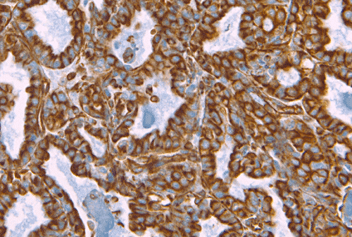
L. S100 |
|
|
|
|
|
||
|
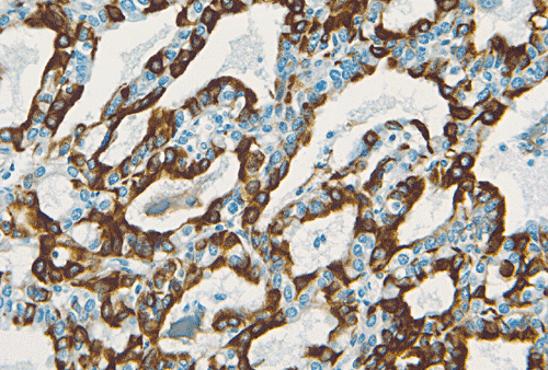
M. Vimentin |
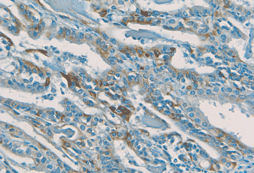
N. AE1/AE3 |
O. GFAP |
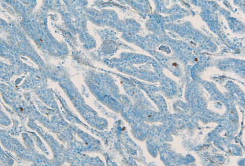
P. Ki67 |
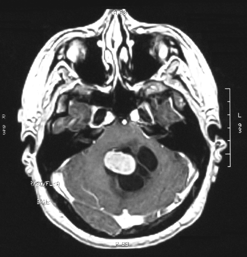
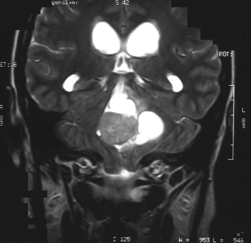
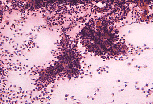
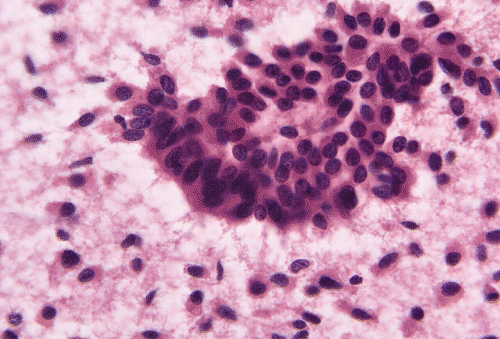
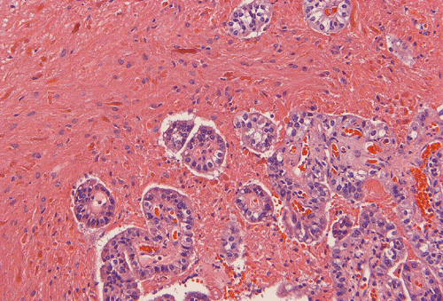
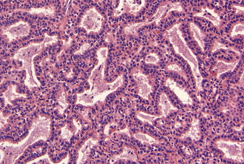
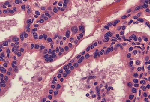
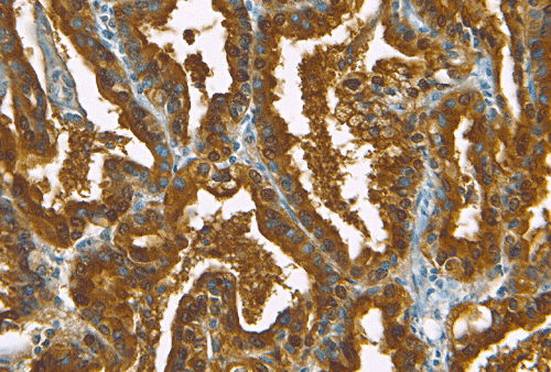
Panel A is T1-weighted image with gadolinium enhancement. Panel B is T2-weighted image. Panel C and D are intraoperative cytologic preparation. Panel E and F are intraoperative frozen sections. Panel G to H are formalin fixed, paraffin embedded sections stained with hematoxylin-eosin stain. Panel L to P are immunohistochemistry for S100 protein, vimentin, cykeratin AE1/AE3, glial fibrillary acidic protein (GFAP) and Ki67 respectively.
Image of the case:
On T1-weighted MR image (Panel A) with gadolinium enhancement, there is a midline cystic lesion with a well-demarcated, oval, mural nodule that has smooth margin. The nodule seems to have a thin and brightly enhancing rim. The core of the nodule shows heterogeneous enhancement. On T2-weighted MR image (Panel B), the nodule is hyperintense to the grey matter and is heterogeneous. No significant edema is demonstrated in the parenchyma surrounding the tumor.
Pathology of the case:
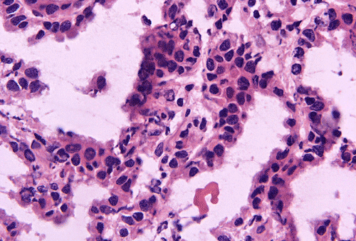
Intraoperative cytologic preparation (Panel C) shows clumps of cells that do not smear out. In between these large clumps are scattered single cells that contain a moderate amount of cytoplasm. On high magnification (Panel D), a papillary pattern is demonstrated in some of the cellular clumps. The nuclei are round to oval, rather monotonous and bland. These cells contain a moderate amount of amphophilic cytoplasm and a well-defined cell membrane.
Intraoperative frozen section (Panel E) show an epithelial neoplasm with the tumor cells arranging in a single layered architecture. There is no necrosis present. The nuclear and cytoplasmic features are in general agreement with the cytologic preparation except that there is a mild degree of nuclear pleomorphism (Panel F).
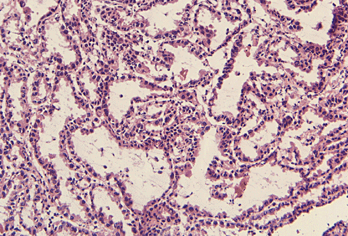
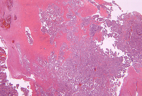
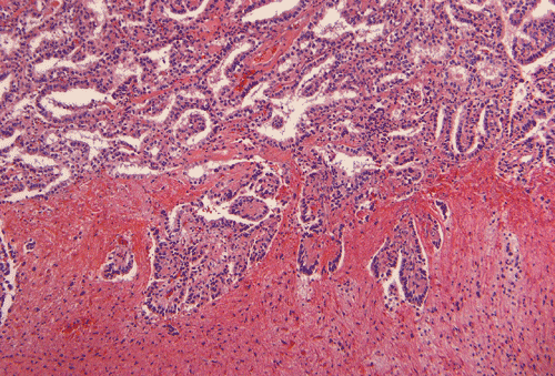
Permanent sections show a papillary epithelial neoplasm with invasion into the brain parenchyma (Panel G, H, and I ). In all areas the tumor cells maintained a single layered architecture. While a papillary structure is present in some area, the tumor cells arrange in a back-to-back cystic architecture intervened by a thin layer of fibrovascular stroma (Panel J). The nuclei are rather bland and monotonous. Mitotic figures are not readily seen (Panel K). On immunohistochemistry, practically all tumor cells strongly express S100 protein (Panel L) and vimentin (Panel M). Most tumor cells are positive for cytokeratin (AE1/AE3) (Panel N). GFAP is expressed by some tumor cells (Panel O). The Ki-67 labeling index is about 1-2% (Panel P).
Comment:
The brain invasion without association with edema that can be demonstrated on T2-weighted MR image is a small surprise. Although the morphologic features are suggestive of choroid plexus papilloma, the possibility of a metastatic low-grade carcinoma has been entertained during intraoperative consultation.
| DIAGNOSIS: Choroid plexus papilloma with brain invasion. |
Discussion: General Information Pathology Prognosis Differential diagnosis
General Information
Choroid plexus papillomas (CPPs) are rare, slow-growing, histologically benign (WHO grade I) intracranial tumors that have the phenotypes of choroids plexus. The incidence is about 0.3 new cases per million population per year and occur more frequently in children than in adults with a mean patient age of 5.2 years. CPPs comprise 0.4-0.6% of brain tumors in patients of all ages and 2-4 % of childhood intracranial tumors. The most common locations are areas where choroids plexus are normally found and, not surprisingly, the ventricles. About 50% occurs in the lateral ventricles, 10% in the third ventricle, CPPs are commonly located in the ventricular system. About half of all cases occur in the lateral ventricles. Most of the rests occur in the fourth ventricle. About 10% occurs in the third ventricles and about 5% involve two or more ventricles. Of all CPPs, 20% occur in patients younger than 1 year of age, and 85% occur in those younger than 10 years of age. About 80% of CPPs occurring in the lateral ventricle are found in patients younger then 20 years old. CPPs occurring in the fourth ventricle are evenly distributed in all age groups and therefore make it the most common location for CPPs that occur in adults as illustrated in our case. The male to female ratio is close to 1:1 for the tumors arising in the lateral ventricles, but 3:2 for tumors arising from the fourth ventricle . CPPs are associated with the Li-Fraumeni cancer syndrome and the Aicardi syndrome 1, 2, 3, 4.
CT scan demonstrates a homogeneously hypodense to slightly hyperdense enhancing mass with or without cyst area and secondary hydrocephalus. High density on CT within the tumor may represent calcification or blood. Homogeneous enhancement on contrast-enhanced CT is typical due to the marked vascularity of these tumors. The appearance of a CPP on MRI is similar to that of a CT scan and shows intermediate-to-strong intensity on both T1- and T2-weighted images 2, 3, 4.
On gross pathologic examination, CPPs appear as lobulated encapsulated masses that are usually attach to the ventricular wall as an exophytic mass that protrude into the ventricle. The mass is well demarcated from the brain parenchyma. Cyst formation and hemorrhage may be seen 2. Microscopically, CPP is featured by papillary structures morphologically resembling the normal choroids plexus and is covered by a single layer of epithelium of different heights (from columnar to flat) supported by a fibrovascular stroma. The epithelial cells are surprisingly regular. Mitotic figures are essentially absent. The cells are usually hobnail shaped and often contain small cytoplasmic granules, no cilia or blepharoplasts. Oncocytic changes and mucin secretion may occur as uncommon histologic features. The similarities with benign papillary mesothelioma are striking. Calcifications, particularly those arising in the posterior fossa, are common. Features of sinister behavior such as multilayered epithelium, high level of pelomorphism, necrosis and mitotic activity are absent. When the specimen is minute and also on frozen sections, it may be difficult to distinguish some truly well-differentiated choroids plexus papilloma from normal choroids plexus. This is usually not a problem in permanent sections.
Although brain invasion as illustrated in this case is an uncommon event, it has been well documented 5. As per one study on CPPs with brain parenchymal invasion in children, these tumors do not appear to behave in a more aggressive fashion then the non-invasive CPPs 5.
Choroid plexus carcinoma is one of the very few carcinomas that have their occurrence limited almost exclusively to children, particularly those under 3 years of age. The carcinomas are from CPP in several aspects. First, there are features of biological malignancy including piling up (multilayered) epithelium with increased pleomorphism, high mitotic activity and necrosis. Second, there is obvious invasion of the adjacent brain parenchyma tissue. Third, there is a loss of the regular papillary architecture of the tumor, at least in the region where invasion is in progress. The tumor cells can arrange in solid sheets to cribriform rather then papillary. The Ki67 labeling in choroid plexus papilloma is 1.9% and in choroid plexus carcinoma is 13.8% 6. In general, the separation between choroids plexus carcinoma and papillomas is usually but not always easy. As they are extremely rare in adult, the possibility of a metastatic carcinoma must be thoroughly entertained before a diagnosis of choroids plexus carcinoma is made in adult.
The term atypical choroids plexus papilloma has been applied to tumors with features in between a CPP and a choroids plexus carcinoma. The diagnostic criteria, however, is not clear cut 2, 6.
Cells of the choroids plexus tumors generally demonstrate ultrastructural features of cells that are involved in fluid transport. Ultrastructural features of choroid plexus papillomas include: the maintenance of the apical-basal polarity of the neoplastic cells; the ocurrence of relatively uncommon cilia and of variable numbers of surface microvilli; the demonstration of a uniform continuous basement membrane outlining the basal plasmalemma; the presence of junctional complexes connecting the cells near the apical surfaces; and the presence of large aggregates of glycogen granules in the infantile and childhood examples.
Immunohistochemically, CPPs are strongly and diffusely positive for the cytokeratins, vimentin, transthyretin and carbonic anhydrase C. Over 90% of cases are positive for S-100. GFAP is negative for the normal choroid plexus whereas is positive in 25-55% of cases 6. Positivity for synaptophysin has also been described. Although transthyretin is rather specific, this protein is also present in the serum and the staining is often accompanied by quite some background for this reason. In addition, its expression tends to diminish in choroids plexus carcinomas. A combined positive staining for cytokeratin, vimentin and S100 would strongly suggest but not be diagnostic of primary choroids plexus tumors. Three out of four cases occurring in the setting of Li-Fraumeni syndrome have a TP53 germline mutation in codon 248. So far no TP53 mutation has been identified in sporadic cases 2.
CPPs can be cure by total resection. In a recent series by McEvoy et al, the 5-year survival rate is 100%, and tumors do not recur in half of the patients who underwent subtotal resection 2, 3, 4. CPPs with a benign cellular appearance but with evidence of local parenchymal invasion still respond to surgical therapy alone, without the need for adjuvant treatment 6, 7.
Differential diagnosis
Choroid plexus papilloma must be differentiated from several other lesions such as villous hypertrophy, choroid plexus carcinoma, papillary ependymoma, metastatic papillary adenocarcinoma, and endolymphatic sac tumor.
Villous hypertrophy is diffuse hypertrophy of the histologically normal choroid plexus in both lateral ventricles. The patients usually have hypersecretory hydrocephalus 2.
The features to distinguish choroids plexus carcinomas from CPPs are mentioned above. In addition, epithelial membrane antigen (EMA) and carcinoembryonic antigen (CEA) are positive in some choroid plexus carcinomas, but are negative in choroid plexus papillomas 1, 2, 6.
Papillary ependymomas do not have a basement membrane, which can be well demonstrated by PAS stain and Jones stain, under the epithelial cells as in CPPs. There are perivascular psuedorosettes with tapering GFAP-positive processes in papillary ependymomas 1. [Click here to see a case of papillary ependymoma]
Endolymphatic sac tumor (low-grade adenocarcinoma probably of endolymphatic origin) is a low-grade adenocarcinoma that is generally seen in adults as a destructive mass in the petrous portion of the temporal bone. They often invavde into the cerebellopontine angle and may extend into the posterior fossa. Immunohistochemically, they are reactive for CEA, EMA and cytokeratin. Histologically, they can be very similar to choroids plexus papilloma. The age of the patient is helpful as choroids plexus tumors are more often seen in childrens. The osseous origin of endolymphatic sac tumor is another important feature. Choroid plexus carcinoma rarely invades extensively into the adjacent bone tissue. [Click here to see a picture of endolymphatic sac tumor]
Reference:
Ironside JW, Moss TH, Louis DN, Lowe JS, Weller RO. Diagnostic Pathology of Nervous System Tumours, Churchill Livingstone, 2002.
Aguzzi A, Brandner and Paulus W. Choroid plexus tumours. In Pathology and Genetics- Tumours of the Nervous System. World Health Organization Classification of Tumours. Kleihues P and Cavenee K. 2000, IARC Press page 84-86.
Islam O and Butt T. Choroid Plexus Papilloma. eMedicine 2005 June. http://www.emedicine.com/radio/topic171.htm
Palmer CA. Choroid Plexus Papilloma. eMedicine 2003 March. http://www.emedicine.com/med/topic2992.htm
Levy ML, Goldfarb A, Hyder DJ, Gonzales-Gomez I, Nelson M, Gilles FH, McComb JG. Choroid plexus tumors in children: significance of stromal invasion. Neurosurgery. 2001 48:303-309.
Rickert CH, Paulus W. Tumors of the choroid plexus. Microsc Res Tech. 2001 52:104-111.
McEvoy AW, Harding BN, Phipps KP, Ellison DW, Elsmore AJ, Thompson D, Harkness W, Hayward RD. Management of choroid plexus tumours in children: 20 years experience at a single neurosurgical centre. Pediatr Neurosurg 2000 32:192-199.
Gyure KA, Morrison AL. Cytokeratin 7 and 20 expression in choroid plexus tumors: utility in differentiating these neoplasms from metastatic carcinomas. Mod Pathol. 2000 13:638-643.
![[Click here to see a picture of endolymphatic sac tumor]](../NeuroTest/Images/SampleQ76.gif)