Trichrome
Iron
| A 38 year-old Woman with a
Subcutaneous Mass in Her Thigh. April, 2005, Case 504-2. Home Page |
Walter F. Bierbaum, M.D., Cheng Z. Liu, M.D., Ph.D. Last update: April 30, 2005.
Department of Pathology, University of Oklahoma Health Sciences Center, Oklahoma City, Oklahoma.
Clinical information: The patient presented with a 3.0 x 3.0 x 1.5 cm subcutaneous mass in the lateral thigh. The mass was excised under the clinical impression of a sebaceous cyst. Macroscopically, the excised specimen is not covered with skin. The following photos are representative photomicrographs of the mass.
|
|
|
|
|
||
| A. | B. | C. | D. | ||
|
|
|
|
|
||
| G. | H. |
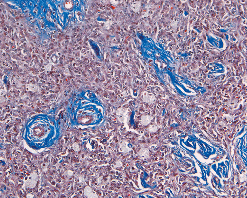
I. Trichrome |
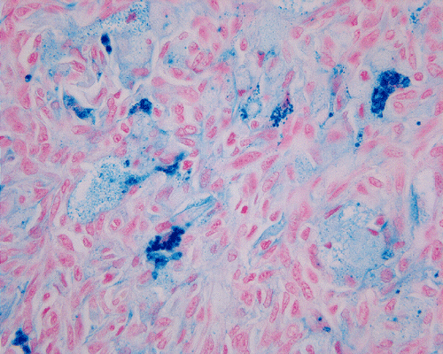
J. Iron |
Pathology of the case:
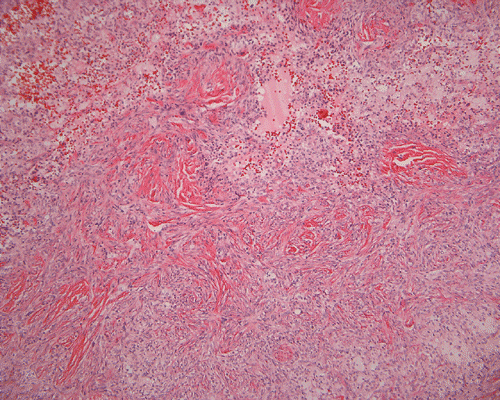
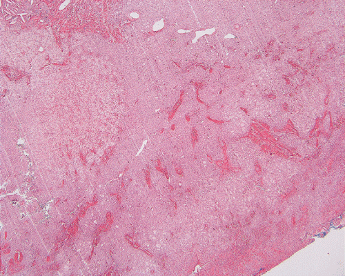
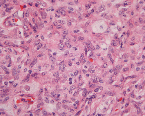
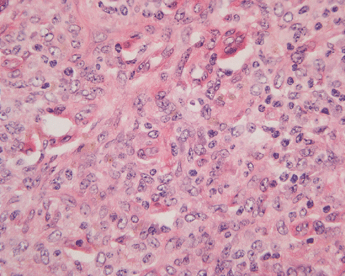
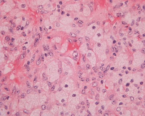
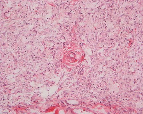
Histologically, the mass has a pushing margin attached with a scant number of skin appendage at one surface of the mass which confirms its superficial location. On low-magnification, the mass has a storiform arrangement of tumor cells that are intermingled with a significant amount of rather broad collagen fibers (Panel A and B). The tumor cells vary from bland, spindle cells with minimal xanthomatous changes (Panel C and D) to polygonal, foamy xanthomatous cells (Panel E and F). The storiform arrangement is more commonly encountered in the periphery and the foamy xanthomatous component is more prominent at the core of this mass. The dense collagen fibers with morphological features comaparable to that of keloid are present and are best demonstrated by Masson's trichrome stain (Panel G). Hemosiderin laden cells can be seen. These cells are far more prominent under the microscope then on the screen. On a histologic stain to demonstrate iron, the depositions are fine and widely distributed. The amount exceeds the expectation based on observation by hematoxylin-eosin stain (Panel H). Giant cells were not readily seen.
| DIAGNOSIS: Lipidized fibrous histiocytoma. |
Discussion: General Information Pathology Differential diagnosis
General Information
Fibrous histiocytomas are common, benign, mesenchymal tumors which typically occur in the lower extremities of young and middle aged adults (women more commonly than men). Most of them have a superficial or subcutaneous location that present as button-like dermal nodules, typically less than 2 cm in greatest diameter. The overlying skin usually has a variable tan to brown color. Tumors with a high content of hemosiderin are more likely to attain a brown color, occasionally mimicking melanoma clinically. Palpation of the lateral aspect of the mass classically produces a central dimple 1. Fibrous histiocytoma are comprised of spindle cells that arrange in a storiform pattern and histiocytes. The histiocytes may harbor hemosiderin or lipid. The periphery of the tumor also typically includes dermal collagen fibers entrapped by the tumor’s spindle cells. A variable mixture of the aforementioned cellular elements (including other cells such as giant cells, lymphocytes, and capillaries) contribute to the remarkable heterogeneity of these tumors. A variety of subtypes with distinctive histologic features have recently been described.
One of the recently described subtypes of benign fibrous histiocytomas is the lipidized fibrous histiocytoma, which is the diagnosis in our case. Iwata et al. recently published a review of 22 cases of this entity 2. This tumor often grows to a slightly larger size than the typical benign fibrous histiocytoma. The median size of the tumors reported by Iwata et al. was 2.5 cm with a maximum size of 8.5 cm. As mentioned in the previous paragraph, benign fibrous histiocytomas are usually less than 2 cm in greatest dimension. In contrast to ordinary fibrous histiocytomas, lipidized fibrous histiocytomas may appear slightly yellow (due to the high lipid content of the tumor) and may have a more exophytic to polypoid appearance. Lipidized fibrous histiocytomas typically occur below the knee and have a particular predilection for the ankle area. These tumors tend to occur in a slightly older population than typical fibrous histiocytoma and are more likely to occur in men (the reverse of fibrous histiocytoma). The age, sex and location of the mass in our patient seem more concordant with ordinary fibrous histiocytomas but the size (3 cm. In greatest dimension) and histologic appearance fit the description of lipidized fibrous histiocytoma. Our tumor, which occurred in the lateral thigh, appeared as a dermal nodule and was initially thought to represent an epidermal inclusion cyst clinically.
Lipidized fibrous histiocytomas are often associated with epidermal thickening. Although the margins are typically well circumscribed, they do not have a capsule. These tumors typically consist of an abundance foamy histiocytes (with mostly bland nuclear features) and characteristic stroma. This stroma contains wire-like collagen fibers of which often display a hyalinized to keloid-like appearance. These two elements (histiocytes and hyalized stroma) occupied greater than 75% of the tumors examined by Iwata et al. A variety of other cells including siderophages and Touton-type giant cells can occur in these tumors. Fibroblasts arranged in a storiform pattern, are usually present, most commonly in the periphery of the tumor. Fifteen of the tumors (from fourteen patients) analyzed by Iwata et al. were stained with immunohistochemical markers. The foams cells in these tumors expressed variable positive staining for CD 68 in all fifteen tumors. CD34 was positive in one tumor. Smooth muscle actin was positive in five tumors 2.
Differential diagnosis
The alternate entity to consider in the differential diagnosis for lipidized fibrous histiocytoma is xanthoma (in particular a variant called plexiform xanthoma). Xanthomas, like lipidized fibrous histiocytomas, tend to contain a large number of lipid-laden histiocytes. The frequent location of xanthomas around the ankle could also trigger some confusion with lipidized fibrous histiocytomas. A feature which leads one to favor a diagnosis of xanthoma is the frequent accumulation of extracellular cholesterol deposits in the form of cholesterol clefts. This feature is usually much less prominent in lipidized fibrous histiocytomas. A histologic feature which favors the diagnosis of lipidized fibrous histiocytomas is the presence of spindle cells in a storiform pattern with entrapped collagen at the periphery, typical features of fibrous histiocytomas. These features should be absent in a xanthoma.
Plexiform xanthoma, a variant of xanthoma, could mimic LFH. This tumor, which frequently involves the dermis, may have a variable mixture of lipid-laden histiocytes and spindle-shaped cells arranged in a storiform pattern. It is differentiated from lipidized fibrous histiocytomas on the basis of its distinct plexiform architecture, frequent and prominent cholesterol clefts, predilection for extensor surfaces, and predominance in male patients 3.
Reference:
Fitzpatrick TB, Johnson RA, Wolff K. Color Atlas and Synopsis of Clinical Dermatology. McGraw-Hill Companies. New York. 1997: 172-173.
Iwata J, Fletcher CD. Lipidized Fibrous Histiocytoma: Clinicopathologic Analysis of 22 cases. Am J Dermatopathol. 2000; 22:126-34.
Michal M, Fanburg-Smith JC. Plexiform Xanthomatous Tumor: A Report of 20 Cases in 12 Patients. Am J Surg Pathol. 2002; 26:1302-11.