Amyloid beta
| A 70 year-old Man with
Confusion and Neurologic Deterioration after a Minor Fall. December, 2005, Case 512-1. Home Page |
Georgeta Varga, M.D.1, Kar-Ming Fung, M.D., Ph.D.2 Last update: December 1, 2005.
1 Department of Neurology and 2 Department of Pathology, University of Oklahoma Health Sciences Center, Oklahoma City, Oklahoma.
Clinical information: The patient was a 70 year-old man with a history of alcoholism, stenosis of the carotid arteries, hypertension, seizure, unstable gait and questionable dementia. He sustained a minor fall about two to three weeks ago and started to have confusion and neurologic deterioration. He died of sepsis about three weeks after the fall. Autopsy was restricted to the brain. Representative images are shown below:
|
|
|
|
|
|
|
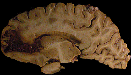
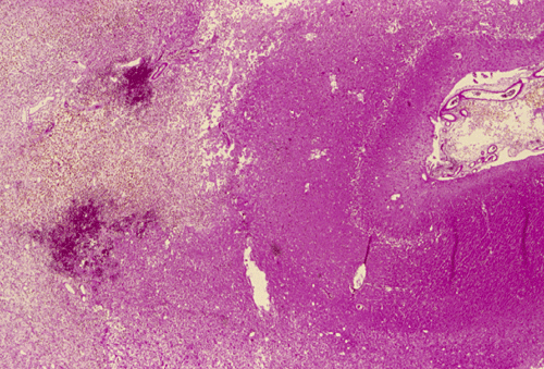
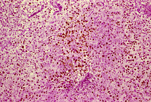
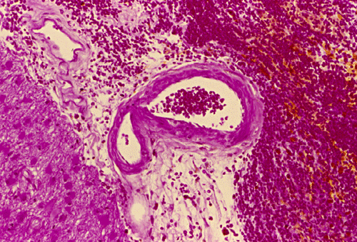
| A. | B. | C. | D. |
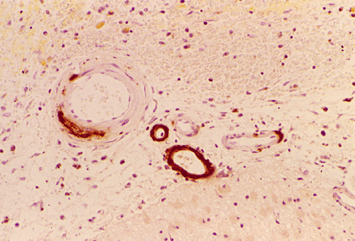
E. Amyloid beta |
Pathology of the case:
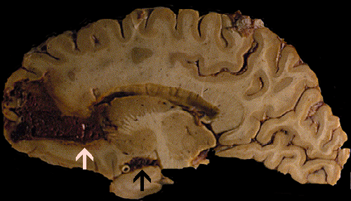
Gross: On external examination of the body, there was no bruising, hemorrhage, or other signs of trauma. The brain weighed 1,300 grams. The cerebellum and brainstem were within normal limits. A large hemorrhage was present in the right frontal lobe (Panel A). The hemorrhage is rimmed by a thin layer of golden brown tissue indicative of hemosiderin deposition that would be produced by old, resolved hemorrhage (White arrow in Panel A). The hemorrhage also extends into the ventricle, without dilating the ventricles, and into the basal subarachnoid space (Black arrow in Panel A). On the left frontal tip and bilateral temporal tips, there were some small areas of golden brown discolorations in the leptomeninges that were consistent with resolved small subarachnoid hemorrhages.
Comment on gross pathology: The golden brown discoloration in the leptomeninges of the temporal tips is probably due to small hemorrhage because of minor falls as the patient has been alcoholic. The question is whether the large hemorrhage related to his fall. The hemorrhage can be produced by trauma but the extent of hemorrhage, however, seems to be out of proportion to a minor trauma as described by the family members of the deceased patients. Since the fall has occurred 2-3 weeks before, the hemosiderin deposition may be resulted from the hemorrhage triggered by that fall. It can also be resulted from prior hemorrhages. The center of the hemorrhage had some spongiotic changes that could suggest a ruptured hemangioma. Other etiologies including coagulopathies, cocaine abuse, vascular malformations and hemangiomas, and vasculopathies were also entertained. The lack of hemorrhagic lesions in other parts of the brain and the body does not support a diagnosis of coagulopathy.
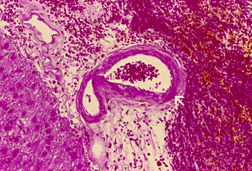
Histopathology: The hemorrhage, not surprisingly, is composed of blood (Panel B). At the periphery of the hemorrhage are areas that are heavily infiltrated by macrophages accompanied by substantial hemosiderin depositions (Panel C). On medium magnification, the blood vessel walls seems to be thickened and some ill-defined deposition are present in the wall (Arrow in Panel D). Immunohistochemistry for amyloid-b (Ab) was performed and the depositions in the vessel wall are positive.
| DIAGNOSIS: Acute hemorrhage secondary to cerebral amyloid angiopathy. |
Pathology of the case:
Discussion: General Information Epidemiology Clinical Pathogenesis Pathology
General Information
Cerebral amyloid angiopathy (CAA) is a very common cause of intracerebral hemorrhage, perhaps only second to hypertensive intracerebral hemorrhage in frequency. They are usually seen in normotensive adults over 60 years of age and occur almost always as cerebral hemorrhage. Hemorhages due to CAA tend to be superficially located and have a distribution distinctly different from those resulted from hypertension. The superficial distribution is mainly because amyloid deposition in CAA is found in the moresmall, superficial vessels and leptomengial vessels. The amyloid can be detected by Congo red, thioflavin S, and immunostaining. The Icelandic and Dutch types of CAA are hereditary. The commonest and sporadic form is due to deposition of (b-amyloid) Ab peptide in the blood vessels. CAA have also been described in some British and Danish families. Deposition of Ab is also a common feature of Alzheimer’s disease, normal aging, and Down’s syndrome.
CAA as an important contributor to stroke in the elderly. Primary hemorrhagic stroke occurs in approximately 100 per 100,000 elderly Americans and Europeans per year or roughly 10% of strokes in this age group. Estimates for the proportion of these hemorrhages that are related to CAA can be obtained from autopsy or clinical series, though each approach has its flaws. Autopsy series, which probably underestimate the true proportion because of the tendency for fewer lobar hemorrhages to come to autopsy,have found 12-15% of hemorrhages in the elderly to be due to CAA , The clinical approach is to count all lobar hemorrhages in the elderly as being due to CAA, which overestimates the disease’s frequency by including some non-CAA entities (e.g. hypertensive hemorrhages that extend to the cortex, vascular malformations). The latter method has yielded estimates of 38-45% (based on series describing patient age and hemorrhage location) 1.
CAA is essentially a vasculopathy feathered by deposition of amyloid in the blood vessel wall particularly in leptomeningeal and cortical arteries, arterioles and, much less frequently, in capillaries and veins. By itself, it constitutes a major risk for hemorrhage in the elderly. This risk is enhanced when there is an underlying hypocoagulation or hemorrhagic tendency such as patients that are treated with anticoagulants. The clinical manifestation is diversified and is dictated by the location of hemorrhage. The patient may have cognitive decline, dementia, stroke, cerebellar and extrapyramidal signs, or a combination of them.
Age represents the strongest risk factor for sporadic CAA-related hemorrhage, as predicted by the steep age-dependence of the underlying pathology. Among 26 CAA patients identified in autopsy series of consecutive hemorrhages all were over age 60 and 23 (88%) over age 70. Similarly, among 95 patients diagnosed with CAA during life by our research group, 93 (98%) were over age 60 at first hemorrhage, 68 (72%) over 70, and 32 (34%) over age 80. Gender does not appear to play an important role in CAA-related hemorrhage. The status of hypertension (HTN) as a risk factor for CAA-related hemorrhage is less clear, in particular the question of whether HTN in conjunction with CAA confers greater risk for lobar hemorrhage than CAA alone 1.
Among potential genetic risk factors for CAA, the apolipoprotein E (APOE) gene has emerged as the strongest predictor of the sporadic form of the .Clinical and pathologic series have suggested both the APOE e2 and e4 alleles as possible risk factors for the presence and earlier onset of CAA-related hemorrhage. The altered APOE allele frequencies appear to be specific for CAA as opposed to hypertensive hemorrhages. Smaller studies have been performed to search for whether mutations associated with familial CAA are also present in patients with sporadic hemorrhages
Though fundamentally a neuropathologic diagnosis, CAA can be identified with good reliability during life by the presence on gradient-echo MRI of multiple, strictly lobar hemorrhages without other definite cause. The relatively good recovery from a first CAA-related hemorrhage is counterbalanced by the tendency for these hemorrhages to recur at approximately 10% per year. Higher rates of recurrence are predicted by a history of previous hemorrhage and the presence of the apolipoprotein E e2 and e4 alleles. Improving tools for the diagnosis and staging of CAA, together with an emerging understanding of Ab deposition and toxicity, form a strong foundation for future trials aimed at prevention of recurring hemorrhages in these patients 1.
CAA-related hemorrhages favor the same cortical or corticosubcortical (“lobar”) regions preferentially affected by the CAA deposits themselves. The acute lobar hemorrhages related to CAA appear to behave like other types of lobar hemorrhage, with symptoms dictated by factors such as hematoma size and location. A slight predilection for frontal and parietal locations has been noted.
Lobar hemorrhages often lobar hemorrhages frequently grow in size during the first hours after onset. They often extend into the subarachnoid space due to their superficial location. Unlike hypertensive hemorrhages. CAA induced lobar hemorrhages only uncommonly rupture into the ventricles. A distinctive feature is their tendency to cause seizures along with the other complications of an acute mass lesion. Non-lobar locations fcr CAA-related hemorrhages are uncommon. Cerebellum contains variable amounts of vascular amyloid and has been reported as an occasional site of hemorrhage. Somewhat surprisingly, primary subarachnoid hemorrhage related to CAA is rare despite extensive pathologic involvement of leptomeningeal vessels.
CAA has been noted in association with several neurologic syndromes other than hemorrhagic stroke. Though fully described only in the last decade, transient neurologic symptoms may be the most common neurologic syndrome related to CAA other than hemorrhagic stroke. Transient symptoms were noted prior to or at presentation in 21 of 95 (22%) consecutive patients at our institution with a diagnosis of CAA and adequate historical information. The symptoms consist of brief (minutes), often stereotyped spells of focal weakness, numbness, paresthesias, or language disturbances; sensory or motor symptoms are often described as spreading across contiguous body parts over seconds to minutes. Although CAA is associated pathologically with ischemic as well as hemorrhagic damage, the transient symptoms and focal seizure may be resulted from small cortical hemorrhages. Supporting this interpretation are the demonstration of small hemorrhages in cortical regions corresponding to the spells, the smooth rather than stepwise spread of the sensory and motor symptoms (atypical for true transient ischemic attacks), and their response to anticonvulsant medication. It is important to differentiate these symptoms from that of true ischemic strokes as treatment with angicoagulants will benefit patients with ischemic stroke but catastrophic for CAA hemorrhage 1.
An uncommon, but potentially treatable clinical presentation of CAA is vasculitis of the central nervous system. Patients with this disorder, like those with central nervous system vasculitis unrelated to CAA, can demonstrate mental status changes, headache, multifocal neurologic deficits, multiple hemorrhagic and nonhemorrhagic lesions (occasionally with a mass-like appearance and inflammatory cells in the cerebrospinal fluid) . Several reported cases responded to immunosuppressive therapy. Vascular-amyloid appears to be the trigger for giant cell reaction and vasculitis in these cases, suggested by the observation of internalized Ab within the giant cells .
The relationship between CAA and dementia is a complex one and probably encompasses a range of different pathogenetic mechanisms. At one end of the spectrum are those relatively infrequent instances in which dementia appear to be a direct result of CAA itself. A number of patients in this category have presented with subacute cognitive decline (over weeks to months) in the setting of a variable combination of seizures, bilateral motor signs, and severe white matter destruction . The observed white matter pathologic change has been ascribed to ischemia from diffuse narrowing of the amyloid-laden cortical penetrating vessels, leading to destructive changes similar to those in subcortical arteriosclerotic leukoencephalopathy (Binswanger’s disease) 1.
Cognitive decline with leukoencephalopathy appears to be most prominent in the Dutch-type familial CAA, where it may precede onset of hemorrhagic strokes.The full-blown syndrome of subacute cognitive decline seems to be uncommon in sporadic CAA, however, appearing in only 1 or 2 of the 95 consecutive patients diagnosed with CAA at our institution.
The more common cause of dementia in sporadic CAA is coexistent Alzheimer disease (AD). AD and CAA have long been observed to overlap at a higher frequency than chance, a likely result of closely related biologies and shared genetic risk factors such as APOE c4 CAA-related hemorrhages were identified in a relatively high proportion (6 of 117, 5.1%) of AD brains in a recent postmortem series . The converse relationship is also present, with pathologic changes of AD appearing in approximately 40-60% of patients with CAA-related hemorrhage. The frequency of clinically manifest dementia (noted prior to first CAA-related hemorrhage) appears somewhat below this range, on the order of 25-40%
Diagnosis
The diagnosis of CAA hemorrhage is not straight forward as amyloid deposition can also occur in Alzheimer’s disease, co-exists with hypertension, and co-exists with other hemorrhagic conditions. As per the Boston criteria, CAA hemorrhage can be defined into four major catetories: definite, probable with supporting pathology, probable, and possible CAA hemorrhage. Brain biopsy provides one mean, although invasive, to confirm the diagnosis. The presence of amyloid in blood vessels is a sensitive marker for CAA-related hemorrhage. Fibrinoid necrosis in the blood vessels, however, is a specific marker for CAA related hemorrhage as they are not seen in autopsy brain with only mild hemorrhage but amyloid deposition in the vessel wall 1, 2.
|
Boston Criteria for the Diagnosis of CAA Hemorrhage1 |
|
1. Definite CAA Hemorrhage Full postmortem examination demonstrating: a. Lobar, cortical, or corticosubcorticai hemorrhage b. Severe CAA with vasculopathy c. Absence of other diagnostic lesion |
|
2. Probable CAA Hemorrhage with Supporting Pathology Clinical data and pathologic tissue (evacuated hematoma or cortical biopsy) demonstrating: a. Lobar, cortical, or corticosubcortical hemorrhage b. Some degree of CAA in specimen c. Absence of other diagnostic lesion |
|
3. Probable CAA Hemorrhage Clinical data and magnetic resonance imaging demonstratIng: a. Multiple hemorrhages restricted to the lobar, cortical, or corticosubcortical region b. Age over 60 c. Absence of other cause of hemorrhage |
|
4. Possible CAA Hemorrhage Clinical data and magnetic resonance imaging demonstrating: a. Single lobar, cortical, or corticosubcortical hemorrhage b. Age over 60 c. Absence of other cause of hemorrhage* |
Amyloidosis falls into a group of disease that shares the common features of distorted or abnormal protein conformation. Other examples of diseases featured by distorted protein conformation include sickle cell disease and prion encephalopathies. A diverse group of proteins including immunoglobulin (in amyloidosis associated with plasmacytic malignancies), b-amyloid (in CAA), and transthyrectin (in amyloid peripheral neuropathy, can assemble into insoluble, b-pleated structures of amyloid. When viewed with Congo red stain under polarized light, they would give an apple-green birefringence.
The most common form is resulted from deposition of Ab peptide, a 4 kD peptide product of cleavage of the amyloid precursor protein (APP), and is closely associated with Alzheimer’s disease. The Icelandic form (hereditary cystatin C amyloid angiopathy) and the Dutch form (hereditary cerebral hemorrhage with amyloidosis-Dutch) are much less frequent, both are transmitted as autosomal dominant traits. Other uncommon types have also been described.
1. Aβ-related cerebral amyloidosis
Multiple mutation sites either within or immediately outside the Alzheimer’s Aβ peptide have been identified in the AβPP gene was found in a condition known as hereditary cerebral hemorrhage with amyloidosis, The Dutch type (hereditary cerebral hemorrhage with amyloidosis-Dutch or HCHWA-D) is resulted from mutation at codon 692 and 693 of APP 3.
2. Cystatin C-related cerebral amyloidosis
Hereditary cerebral hemorrhage with amyloidosis, Icelandic type (HCHWA-I) is an autosomal dominant disorder characterized by massive amyloid deposition within small arteries and arterioles of leptomeninges, cerebral cortex, basal ganglia, brainstem and cerebellum. The Icelandic form (hereditary cystatin C amyloid angiopathy) is resulted from single nucletide substitution at codon 68 of cystatin C leading to replacement of glutamine by leucine. Patients also have abnormally low level of cystin in the cerebral spinal fluid 3.
3. ABri-related cerebral amyloidosis
Familial British dementia, is an early onset autosomal dominant disorder clinically characterized by progressive dementia, spastic tetraparesis and cerebellar ataxia. Severe amyloid angiopathy of the brain and spinal cord with perivascular amyloid plaque formation, parenchymal plaques affecting limbic aras, cerebellum, and occasionally cerebral cortex, neurofibrillary degeneration f hippocampal neurons as well as periventricular white matter changes are the main neuropathological hallmarks of the disease 3.
4. ADan-related cerebral amyloidosis
Familial Danish dementia, also known as heredopathia ophthalmo-oto-encephalacia, is an early-onset autosomal dominant disorder originating in the Djursland peninsula, Denmark. The disease, identified in nine cases spanning three generations of a single family, is clinically characterized by the development of cataracts, deafness, progressive ataxia and dementia. Cataracts seem to be the early manifestation of the disease, starting before the age of 30, whereas impaired hearing usually develops 10-20 years later. Cerebellar ataxia occurs shortly after the ge of 40, followed by paranoid psychosis and dementia 10 years later. Most patients die in their fifth to sixth decade of life. The disease is neuropathologically characterized by diffuse brain atrophy with a particularly severe involvement of the cerebellum, cerebral cortex and white matter, as well as by the presence of very thin and almost demyelinated cranial nerves. There is a widespread amyloid angiopathy in the blood vessels of the cerebrum, choroid plexus, cerebellum, spinal cord, and retina. The presence of parenchymal plaques and neurofibrillary tangles is the major histological finding in the hippocampus, whereas the cerebral white matter also shows some ischemic lesions 3.
5. Transthyretin related amyloidosis
Transthyretin related amyloidosis is usually associated with peripheral neuropathy due to amyloidosis. Leptomeningeal amyloid deposits in transthyretin amyloidosis have been reported a number of times in subjects with various transthyretin mutations. The recognition of CNS dysfunction in transthyretin amyloidosis is important. While the liver may be the origin of most or all of transthyretin in the circulating blood plasma, transthyretin in the cerebral spinal fluid is synthesized by cells of the choroid plexus, and vitreous transthyretin probably by the retinal pigment epithelium 3.
As we have mentioned before that CAA has close ties with Alzheimer's disease, some features of Alzheimer's disease such as neurotic plaques and neurofibrillary tangles can also be found in some variants of the CAAs and they are summarized as follows 3:
|
|
CAA |
Diffuse plaques |
Neuritic plaques |
Neurofibrillary Tangles |
|
Icelandic type |
● |
|
|
|
|
Dutch type |
● |
● |
|
|
|
Familial Danish dementia & Familial British dementia |
● |
● |
● |
● |
|
Alzheimer's disease |
● |
● |
● |
● |
Grossly, the smaller hemorrhage tends to be superficial rather then deep. For large hemorrhages that have deep extension, it may not be possible to tell whether the epicenter of hemorrhage is superficial or deep. Their superficial location tends to make a small amount of subarachnoid hemorrhage. Extension into the ventricle is uncommon. Typically, small hemorrhage at different stages of resolution is present. The hemorrhages are found as lobar hemorrhage in the white matter in contrast to the hemorrhages that are found in hypertension and are often found in the basal ganglia. CAA hemorrhage is almost always cerebral with the brainstem and cerebellum rarely affected. This is an interesting phenomenon as the cerebellar folia have a large surface area and numerous leptomeningeal blood vessels. CAA hemorrhagedoes not appear to have a predilection to occur in leptomeningeal vessels although these vessels typically contains amyloid deposition in the wall.
Microscopically, the pathology of hemorrhage is not different from that of hemorrhage due to other causes. However, the blood vessels, particular the arterioles and the leptomeningeal vessels appear to have thickened wall and some clumpy irregular, eosinophilic depositions may be seen. The depositions are also positive for periodic acid Schiff stain. The affected vessels in Ab caused CAA often have segmental dilatations, microaneurysm formation, and fibrinoid necrosis. The small muscle layer is often destroyed. In severely affected vessels, a double-barrel vessel wall is present.The amyloid depositions will stain bright orange-red with Congo red and will give a green birefrigence under polarized light. In addition, immunohistochemical detection would be positive for Ab in many of the sporadic cases. P-component which almost always co-deposit with amyloid can also be detected by immunohistochemistry. At the ultrastructural level, Ab amyloid appears as clumps and bundles of straight filaments of 10 nm in diameter.
Reference:
Greenberg SM. Clinical aspects and diagnostic criteria of sporadic CAA-related hemorrhage. In Cerebral Amyloid Angiopathy in Alzheimer ‘s Disease and Related Disorders. Verbeek MM, de Waal RMW, Vinters HV (eds), Kluwer Academic Publishers, Dordrecht, 2000, Chapter 1, pp. 3-19,
Frangione B, Revesz T, Vidal R, Holton J, Lashley T, Houlden H, Wood N, Rostagno A, Plant G, Ghiso J. Familial cerebral amyloid angiopathy related to stroke and dementia. Amyloid. 2001 Suppl 1:36-42.