| A 22 year-old Man with
Metastatic Mature Testicular Teratoma in Lymph Node. January, 2006, Case 601-2. Home Page |
Kar-Ming Fung, M.D., Ph.D.1, Alberto G. Ayala, M.D.2, Jae Y. Ro, M.D., Ph.D.2
1 Department of Pathology, University of Oklahoma Health Science Center, Oklahoma City, OK, 2 Weill Cornell University and The Methodist Hospital Houston, TX.. Last update December 31, 2005
Clinical information:
A 22 year old man presented to the hospital with abdominal pain, nausea and vomiting. Physical examination disclosed right supraclavicular adenopathy and a left testicular mass. His beta-HCG in serum was 394,000 mIU/ml and alpha-fetoprotein in serum was 2,100 ng/dL. A CT scan disclosed multiple complex heterogeneous masses involving the mediastinum, chest, retroperitoneum, lesser sac and mesentery of the abdomen, and possible pelvic lymph nodes. An inguinal radical orchiectomy was performed and pathologic examination revealed a mixed germ cell tumor of the testis that was composed of 70% teratoma and 30% seminoma. He was treated with chemotherapy consisting of BEP (bleomycin, etoposide and cisplatin).
His tumor markers normalized five months after treatment.. Multiple large masses in the mediastinum, retroperitoneum, and abdomen, however, could still be demonstrated by CT scan. Biopsy material of a supraclavicular lymph node revealed only mature teratomatous elements. He was treated with chemotherapy consisting of VIP (vinblastine, ifosfamide and cisplatin). Although there was a brief period of elevated beta-HCG level in serum before the chemotherapy, his beta-HCG and alpha-fetoprotein levels were within normal limits 10 months after orchiectomy and chemotherapy.
An extensive lymphadenectomy was performed and the followings are representative photomicrographs:
|
|
|
|
|
||
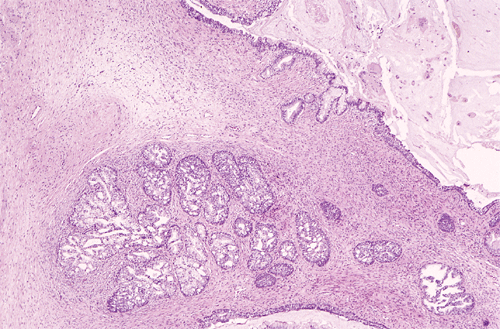
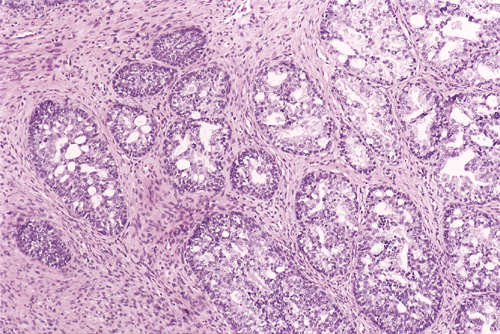
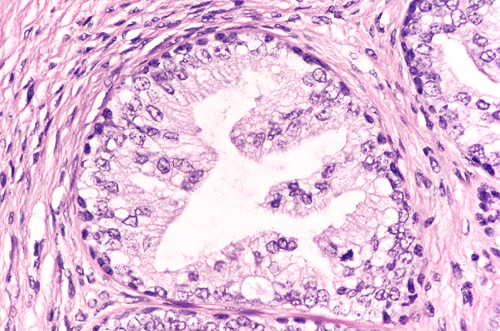
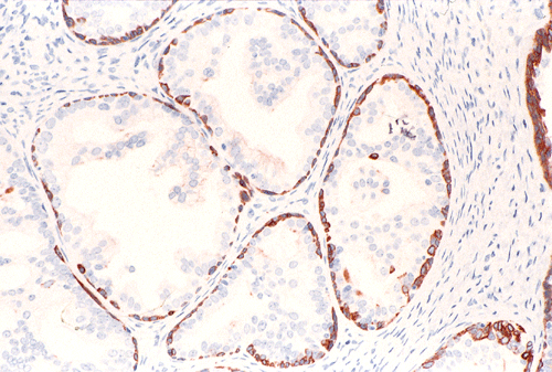
| A. | B. | C. | D. |
Pathology of the case:
An extensive lymphadenectomy of the chest and abdomen was performed and yielded multiple masses of enlarged and matted lymph nodes with the largest one measruing12 x 6 x 3 cm. While some of the lymph nodes were completely necrotic, others contained viable tissue.
Histologic examination revealed complete replacement of lymph nodes by neoplastic tissue composed of mature mesenchymal, epithelial, and neural components associated with extensive fibrosis. A substantial amount of the tumor was composed of benign fibroblastic growth decorated by nests of epithelium with cyst formation. While most of the epithelial elements are that of intestinal or respiratory type (Panel A, arrow), a few small foci (less than 5%) appeared distinctly different. These foci consisted of small epithelial acini with a lobulo- centric arrangement (Panel A). The acini were composed of two cell types: cuboidal epithelial cells lining the lumens and smaller, more hyperchromatic cells located at the base of the acini. The epithelial cells showed proliferative features such as papillary luminal projections without fibrovascular cores, intraluminal bridging of these projections and mounds of pseudostratified cells (Panel B). These cells had abundant clear and granular cytoplasm. The nuclei were enlarged compared to the stromal cells, had fine chromatin pattern and occasionally had prominent nucleoli (Panel C). High molecular weight cytokeratin (34BE12) was expressed by the cells located at the periphery of these acini as a continuous or interrupted layer (Panel D). No immature teratoma elements or residual seminoma were noted in any of the lymph nodes.
Immunohistochemistry also disclosed strong expression of prostate specific antigen (PSA) and prostate specific acid phosphatase (PSAP) in the glandular elements. Prostatic lineage of these cells was thus confirmed. The histologic and immunohistochemical features of these epithelial cell nests were those of a high grade prostatic intraepithelial neoplasia (PIN). The differential diagnosis in this case included benign and neoplastic prostate tissue as well as other glandular tissues in mature teratoma. The morphology and immunohistochemical features, however, were diagnostic for a high grade PIN. There was no invasive adenocarcinoma of prostate in the metastatic mature teratoma.
| DIAGNOSIS: High-grade prostatic intraepithelial neoplasm (PIN) in metastasizing mature teratoma. |
Discussion: General Information Pathology
General Information
Mixed germ cell tumors of testis that present with distant metastasis and elevated serum markers such as HCG and α-fetoprotein are generally treated chemotherapy with or without orchiectomy. When residual masses are resected after the chemotherapy, generally necrosis or fibrosis is what remains in the specimen. Necrosis most likely represents highly proliferative components like embryonal carcinoma and endodermal sinus tumor and possibly seminoma that are affected by the chemotherapy. It is not unusual to find only mature teratoma as the viable tumor in these specimens, as seen in this case, since mature elements are not affected by the chemotherapeutic agents. The role of the pathologists is to identify residual high-grade elements such as embryonal carcinoma and/or endodermal sinus tumor since the presence of such elements mitigates for further aggressive chemotherapy.
Pure mature teratoma is very rare after puberty 1. In children younger than 5 years of age it is considered benign. In adults, on the other hand, 16.7 % of the clinical stage A cases metastasize to retroperitoneal lymph nodes 2.
The differentiated elements in teratomas vary according to their site of origin 3. Tissue with prostatic differentiation is extremely rare. Although it has been dected in 4 in ovarian teratomas, it has not been described in testicular mature teratomas. However, there was no well documented cases in testicular mature teratomas. Benign and malignant tumors other than germ cell tumors can arise in association with teratomas. While squamous cell carcinoma 5 is the most common malignant transformation, other tumors such as glomus tumor 6, melanoma 7 small cell carcinoma 8, and adenocarcinoma 9 have been described in mature teratomas. A study of squamous cell carcinoma in situ in ovarian teratomas 10 suggests that some of these malignant tumors may arise from incipient preneoplastic lesions. Presence of high grade prostatic intraepithelial neoplasia or prostatic adenocarcinoma in mature teratoma has not been described and therfore, their biological significance is uncertain.
In summary, to our knowledge this is the first case showing high grade PIN morphology in prostatic tissue arising in a metastasizing mature teratoma being reported in the English literature.
Reference:
Ulbright TM. Germ cell neoplasms of the testis. Am J Surg Pathol 1993; 17:1075-91.
Leibovitch I, Foster RS, Ulbright TM, Donohue JP. Adult primary pure teratoma of the testis. The Indiana experience. Cancer 1995; 75; 2244-50.
Murphy WM. Urological pathology. Pp 369 2nd edition. 1997 WB Saunders.
Vadmal M, Hajdu SI. Prostatic tissue in benign cystic ovarian teratomas. Hum Pathol 1996; 27:428-9.
Noumoff JS, LiVolsi VA, Deger RB, Montone KT, Faruqi SA. Chromosome analysis and comparison of the benign cystic and malignant squamous component of an ovarian teratoma. Cancer Genet Cytogenet 2001; 125:59-62.
Silver SA, Tavassoli FA. Glomus tumor arising in a mature teratoma of the ovary: report of a case simulating a metastasis from cervical squamous carcinoma. Arch Pathol Lab Med 2000;124:1373-5.
Vigliani R, Iandolo M, Lacivita A. Mature ovarian cystic teratoma with combined squamous cell carcinoma and malignant melanoma. Virchows Arch 1998; 433:381-4.
Lim SC, Choi SJ, Suh CH. A case of small cell carcinoma arising in a mature cystic teratoma of the ovary. Pathol Int 1998; 48:834-9.
Ng EW, Porcu P, Loehrer PJ Sr. Sacrococcygeal teratoma in adults: case reports and a review of the literature. Cancer 1999; 86:1198-202.
Tobon H, Surti U, Naus GJ, Hoffner L, Hemphill RW. Squamous cell carcinoma in situ arising in an ovarian mature cystic teratoma. Report of one case with histopathologic, cytogenetic, and flow cytometric DNA content analysis. Arch Pathol Lab Med 1991 115:172-4.