| A premature neonate (30
weeks of gestation) with Feeding Problems and Abdominal Distension. February, 2006, Case 6012-1. Home Page |
Alde Carlo P. Gavino, M.D., W. Frederick Bierbaum, M.D., Willard Aronson, M.D. Last update March 31, 2006.
Department of Pathology, University of Oklahoma Health Sciences Center, Oklahoma City, OK
Clinical information: The patient was a 3-week 5-day old baby boy born at the 30th week of gestation to a 27 year-old mother. The pregnancy was complicated by maternal ethanol use and prolonged premature rupture of membranes. The patient’s APGAR scores were 2 at 1 minute, 6 at 5 minutes, and 8 at 10 minutes. After delivery, the patient was admitted to the intensive care unit for prematurity, possible sepsis, and respiratory distress. Blood cultures in the first week of life were negative. He was weaned off respiratory support shortly after the first week of life. Two weeks after birth, the patient began having some feeding intolerance with high residuals by gavage feeds. A few days later, he developed abdominal distention. An abdominal radiograph revealed free air, consistent with intestinal perforation. A Penrose drain was placed. The patient also had a tonic-clonic seizure at one point and then started having oxygen desaturations. His clinical condition deteriorated with metabolic acidosis, hypotension, cardiac arrhythmia, bradycardia, and hypoperfusion despite aggressive clinical treatments that included the use of vasopressors. He eventually succumbed to these multiple medical problems.
An autopsy was performed. The following are representative images of the gastrointestinal tract from the autopsy:
|
|
|
|
|
|
|
| A. | B. | C. | D. | E. | |
|
|
|
|
|
||
| F. | G. | H. | I. |
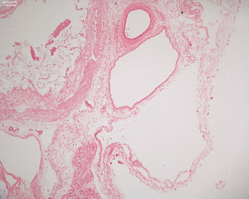
Pathology of the case:
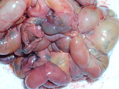
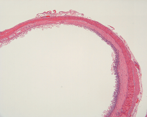
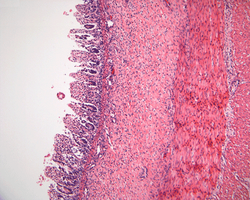
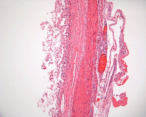
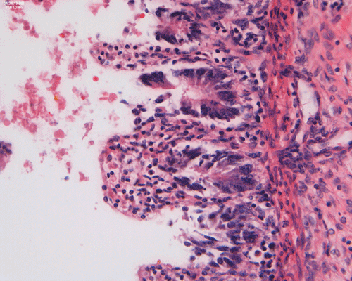
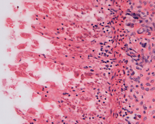
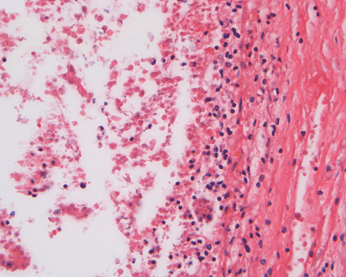
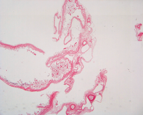
Discussion of the pathology is limited to the alimentary canal. On autopsy, the intestinal loops are largely dilated with gas accumulation. On closer examination, many small, subserosal bubbles can be seen (arrows in Panel A). The intestinal wall is also thinned out as a result of the dilatation by gas (Panel B). In the less affected areas, the intestinal wall is not thinned out and the mucosa can still be recognized as largely intact (with autolytic changes) (Panel C and E). In the thinned out areas, the mucosa appear pink and necrotic (Panel D, F and G). The subserosal bubbles appear to be empty vesicles filled with air and they are found most frequently in the most necrotic region. In general, these features indicate widespread infarction with gas production (Panel H and I).
| DIAGNOSIS: Necrotizing enterocolitis. |
Discussion: Introduction Pathophysiology Clinical Diagnosis Pathology
Necrotizing enterocolitis (NEC) is a life-threatening disease of the newborn characterized by varying degrees of inflammation and coagulative and hemorrhagic mucosal or transmucosal intestinal necrosis. NEC typically affects premature infants with birth weights ranging from 1000 to 1500 grams. These patients are usually ill with respiratory distress syndrome. The average age of onset is on the first week of life, although the disease may develop as early as the first day after birth. Term infants have also been occasionally affected. The incidence of NEC ranges from 1 to 5% of admissions to neonatal intensive care units. Because of improved management of premature infants, the incidence seems to be rising recently. There is no racial or sexual predilection.
Management of NEC includes cessation of oral feedings, administration of antibiotics, and surgery for perforation or other evidence of severe bowel compromise. Lengthy intestinal resection may result in short bowel syndrome. Long term parenteral nutrition is required in many cases. The overall mortality rate from NEC has been estimated to be about 20-30% and is even higher in lower-birth-weight infants.
Several factors have been implicated to predispose to the development of NEC. They include intestinal ischemia, bacterial colonization, oral milk feedings, and premature birth. Intestinal ischemia may be brought about by a variety of conditions such as reduced splanchnic perfusion, systemic hypoperfusion, systemic hypoxia, or local factors such as intestinal gaseous distention. Bacterial colonization is nearly always present in NEC. Organisms recovered nonspecifically from cultures have included Clostridium difficile, Clostridium perfringens, Escherichia coli, and Staphylococcus epidermidis. Secretion of inflammatory mediators such as platelet activating factor and tumor necrosis factor is induced by the presence of bacterial toxins. These mediators are important in the development of the necrotic process that occurs in NEC. NEC, however, is traditionally not considered to be an infectious process in that no specific organisms or group of organisms have been implicated to cause the disease. Milk formulas are thought to cause gut flora to proliferate and may affect intestinal perfusion. Other conditions that have been shown to be associated with the causation of NEC include polycythemia, administration of hypertonic milk or medicines, and rapid feeding protocols. The final common pathway is mucosal injury and subsequent infection leading to bowel necrosis.
Clinical Manifestations
The first signs of NEC are abdominal distention and gastric retention of feedings. The onset of the disease is often insidious. Symptoms vary from mild, with only guaiac-positive stools, to severe, with peritonitis, bowel perforation, shock, apnea, and death. Bloody stools are manifested in 25% of patients. One-third of patients have a fulminant course with intestinal perforation, and a similar number have bacterial sepsis. Progression of symptoms may be rapid but it is unusual for the disease to progress from mild to severe after 72 hours.Complications of NEC include stricture formation and bowel perforation. Strictures form secondary to progressive circumferential submucosal fibrosis (if the bowel affected is not resected during the acute phase). Ten to 20% of infants between 3 and 10 or more weeks after NEC has been diagnosed develop strictures. Perforation leads to peritonitis. Other complications include sepsis and compromised nutrition.
A very high index of suspicion is essential in diagnosing NEC. A suggestive clinical picture coupled with radiographic evidence of pneumatosis intestinalis or gas in the portal or hepatic veins is virtually diagnostic. NEC lends itself diagnosable by non-invasive methods because pneumatosis intestinalis is demonstrable by plain abdominal radiography. By the time treatment is started, 50 -75% of patients already have pneumatosis. Portal vein gas is a sign of severe disease and pneumoperitoneum indicates perforation. It must be emphasized, however, that positive radiologic signs may be absent in one-third of NEC patients, in which case, the diagnosis is confirmed at surgery or autopsy.
In the majority of cases (about 80%) of NEC, the terminal ileum and cecum and the ascending colon are the most severely affected bowel segments. The disease process might be restricted to the small intestines or colon alone or it may diffusely involve the bowels. Half of the cases present with patchy segmental necrosis of the bowels with intervening spared areas while the rest present with continuous and circumferential necrosis, dilatation, and friability.
Coagulative and hemorrhagic necrosis is the key pathologic feature of NEC. Necrosis is limited to the mucosa in the early stages but at least focally transmural in specimens removed from surgical resection or at autopsy. In 10% of cases, focal necrotic pseudomembrane formation is seen.
A mixed acute and chronic inflammatory infiltrate is commonly found, limited to the mucosa in some foci but transmural in others. Inflammation and necrosis often occur together in a given segment, but in some instances, one or the other may predominate.
Pneumatosis intestinalis, usually limited to the submucosa, is seen in 50% of surgical specimens with NEC. The gas bubbles have been shown to contain hydrogen, a product of bacterial fermentation. More than 50% of patients who undergo laparotomy show focal reparative epithelial changes and other evidence of healing, such as the formation of granulation tissue and crypt distortion. Villous atrophy may also be observed. Such reparative changes suggest that NEC evolves gradually before a catastrophic event, such as perforation, brings it to clinical attention.
A mixed intestinal flora is often visible in the lumen or within the necrotic superficial mucosa. Fungal growth has been found in 3.5% of cases in one large series. Occasional epidemics with Escherichia coli, Klebsiella pneumoniae, Clostridium difficile, and rotavirus have been reported. However, in neither sporadic cases nor in most epidemics is a specific pathogen found.