
AE1/AE3
AE1/AE3
Vimentin
| A 11 year-old Boy with a
Tibial Mass. March, 2006, Case 803-2. Home Page |
Walter F. Bierbaum, M.D., Jian T. Yang, M.D. Ph.D. Last update: May 31, 2006.
Department of Pathology, University of Oklahoma Health Sciences Center, Oklahoma City, Oklahoma
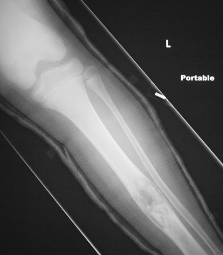
Clinical information: The patient is an 12 year old boy who presented to the emergency room with pain and swelling of the left leg. He first experienced the symptoms while practicing football. An X-ray was performed (Panel A) revealing a pathologic fracture through the shaft of the left tibia. The fracture was associated with a large soap bubble-like lesion in the middle diaphysis. A curettage biopsy was performed.
|
|
|
|
|
|
|
| A. | B. | C. | D. | E. | |
|
|
|
|
|
|
|
| F. | G. | I. | I. | J. | |
|
|
|
|
|||
|
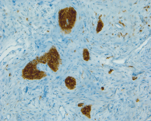
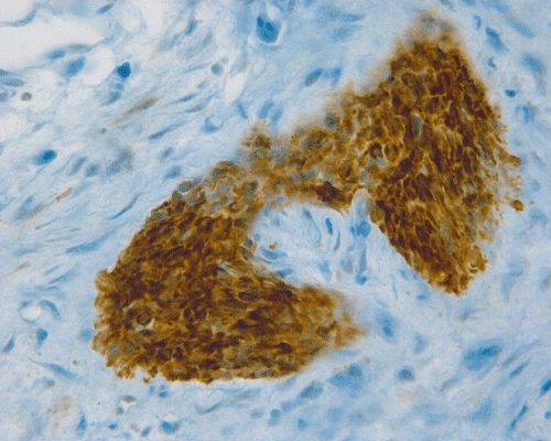
K. AE1/AE3 |
L. AE1/AE3 |
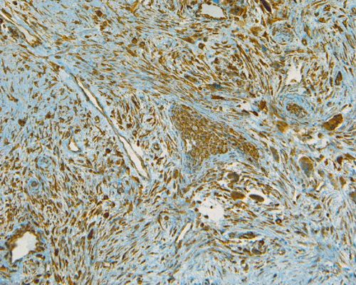
M. Vimentin |
| DIAGNOSIS: Adamantinoma, classic type with pathologic fracture. |
Radiology of the case: On the plain film, there is an osteolytic lesion (Panel A) associated with a fracture (fracture line is illustrated by the black arrow). The lesion is predominantly intracortical. However, the cortex is thinned out significantly in some areas and is breached (white arrow). The medullary cavity is also involved.
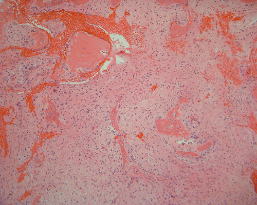
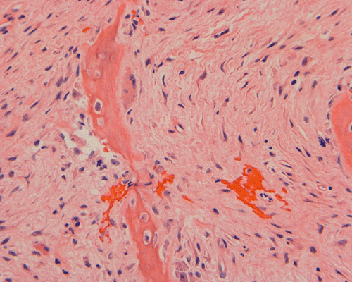
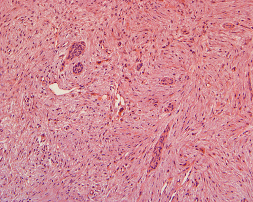
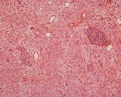
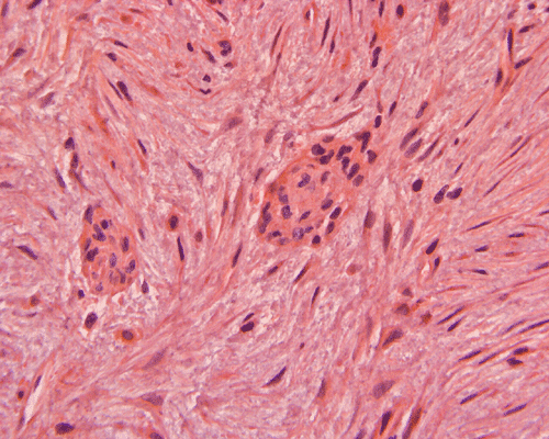
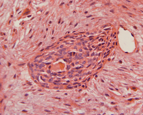
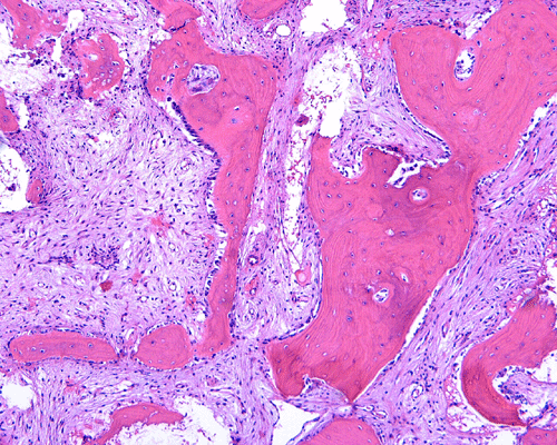
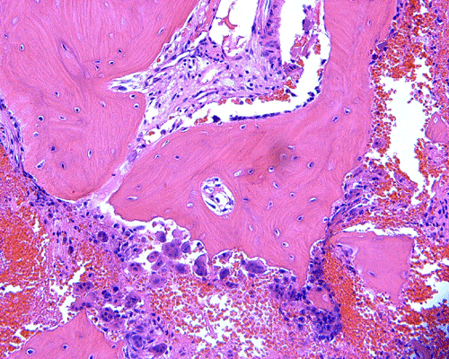
Pathology of the case: The specimen consists of multiple small pieces of irregular tissue fragments obtained by curretage. Histologically, the lesion tumor consists of bland spindled cells embedded in a collagenous matrix and arranged in a storiform pattern with frequent hemorrhage (Panel B). Entrapped residual bone associated with foci of osteoclastic type giant cells are present (Panel C). A scant number of epithelial islands are also scattered within this spindle cell background (Panel D, E, and F). On high magnification, no significant nuclear atypia or pleomorphism is noted in either the spindle cell or epithelial cell components. In other parts of the specimen, the spindle cell tumor is admixed with spiculated fragments of woven bone rimmed by osteoblasts (Panel H, I, and J). Small islandsIt of epithelial cells are also noted in these areas (Arrow in Panel H).
On immunohistochemistry, the epithelial islands are positive for cytokeratin AE1 and AE3 (Panel K and L). Both the spindle cell component and epithelial cells are positive for vimentin (Panel M).
Comment: Although the age and histological pattern of this case is highly compatible with the differentiated type of adamantinoma, there is extension of the tumor into the medullary cavity (see discussion below). For the later feature, this case is more likely a classic adamantinoma with prominent osteofibrous dysplasia-like areas. Our outside consultant concurred with our diagnosis. The prognosis for classic adamantinoma is worse than differentiated adamantinoma. The classic variety is more likely to recur following surgery and is much more likely to metastasize. Intralesional or marginal excision may be the most significant risk factor for a local recurrence or metastasis. The lung is the most common site for metastasis. The treatment for differentiated adamantinoma is much less radical, consisting of a more limited en bloc cortical excision.
Discussion: General Information Pathology Differential Diagnosis
General Information
Adamantinomas can be separated into two distinct groups, classic and differentiated. Each group manifests distinct clinical radiological and histologic findings 1, 2.
The classic form of adamantinoma is a rare tumor that typically occurs within the tibia or fibula of patients in the 3rd to 7th decade of life. Radiographically, the tumor may be intracortical, intramedullary, or involving both cortical and medullary cavity with extension to the adjacent soft tissue.Some tumors have both features. Their radiologic features are hightly distinctive and often diagnostic. Most commonly they are seen in the midshaft of the tibia. The proximal or distal tibial region is the next most commonly affected sites. Histologically, they are very similar to the odontogenic adamantinoma of the jaw bones such as ameloblastoma 2.
Differentiated (osteofibrous dysplasia-like) adamantinoma is also a rare tumor. In contrast to the classic form, it is seen most commonly in the1st to 2nd decade of life. It is exclusively intracortical in the tibia with frequent synchronous involvement of the ipsilateral fibula. Histologically, it is characterized by small nests of epithelial neoplasic cells scattered individual epithelial cells, or both with an oseofibrous dysplasia-like background. Their radiologic appearance is identical to osteofibrous dysplasia, consisting of radiolucent areas surrounded by a rim of intact cortical bone. In contrast to classic adamantinoma, the differentiated variety does not extend either into the medullary cavity or outside of the periosteum into surrounding soft tissue. Therefore, this entity has radiographic, histopathologic and clinical features overalpping with osteofibrous dysplasia 2.
The classic type of adamantinomas of the long bone are low grade malignant tumors that are slow growing, locally destructive. They have a high recurrence rate and about 25% of them will metastasize, usually late metastases and most commonly to the lung. As per the description of Czerniak and Dorfman, no differentiated (osteofibrous dysplasia-like) adamantinoma has metastasized in their experience 2, 3.
Classic adamantinomas are often intracortical and may extend beyond the periosteum into surrounding soft tissue. Its histologic features recapitulate those of the synonymous tumor of the jaw. The epithelial component of these tumors is usually prominent. The most common epithelial patterns include basaloid, spindled, tubular, and squamoid. An intervening osteofibrous dysplasia-like pattern can also be seen in classic adamantinoma, but is usually focal, occupying the periphery of the tumor.
Differentiated (osteofibrous dysplasia-like) adamantinomas are exclusively intracortical. The microscopic appearance of differentiated adamantinoma resembles osteofibrous dysplasia. Spindled cells with a storiform pattern mingle with fragments of bone. The bony fragments are typically rimmed by osteoblasts and exhibit gradual progression from woven to lamellar bone as one moves from the center to the periphery of the lesion. Epithelial islands can be seen interspersed focally within the aforementioned osteofibrous dysplasia-like stroma. The epithelial component is much less prominent than in the classic variety of adamantinoma. Immunohistochemistry for cytokeratin is a very useful adjunct to demonstrate these scattered epithelial elements and they can be very small and subtle as demonstrated in this case.
Differential Diagnosis
The distinction between osteofibrous dysplasia and long bone adamantinoma can be difficult, particularly in small biopsy specimens. In general, radiographic features favoring classic adamantinoma over osteofibrous dysplasia include a multilayered periosteal reaction and moth-eaten bony destruction 4. In contrast, differentiated adamantinoma and osteofibrous dysplasia are frequently radiologically identical, and have analogous histologic appearances. One distinguishes the two by noticing the epithelial islands within differentiated adamantinoma. Immunohistochemical stains, most commonly cytokeratin, can help demonstrate the diagnostic epithelial areas. Of note, a peripheral biopsy of classic adamantinoma may also have a relatively high proportion of the osteofibrous-like stroma (as we noticed in this case). Like the differentiated form of adamintoma, a diagnosis can be established by recognition of epithelial nests within the tumor.
References:
WHO Classification of Tumors: Tumors of Soft Tissue and Bone. IARC Press. Lyon. 2002: 332-334, 343-344.
Dorfman H, Czerniak B. Bone Tumors. C.V. Moseby. 1998: 949-973.
Hazelbag HM, Taminiau AH, Fleuren GJ, Hogendoorn PC. Adamantinoma of the long bones. A clinicopathological study of thirty-two patients with emphasis on histological subtype, precursor lesion, and biological behavior. J Bone Joint Surg Am. 1994 76(:1482-99.
Bloem JL, van der Heul RO, Schuttevaer HM, Kuipers D. Fibrous dysplasia vs adamantinoma of the tibia: differentiation based on discriminant analysis of clinical and plain film findings. AJR Am J Roentgenol. 1991 156:1017-23.
Cases of the Month Evaluation Coordinator: KarMing-Fung@ouhsc.edu