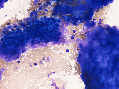
DiffQuick
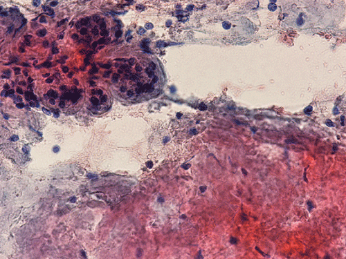
PAP stain
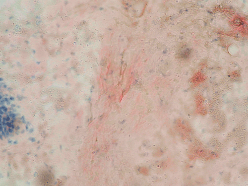
Congo red
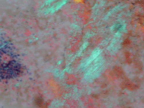
Congo red polarized
Congo red
Congo red polarized
| A 54 year-old Man with
Bilateral Enlargement of Submandibular Glands and Proteinuria. April, 2006, Case 604-2. Home Page |
Pablo Souza, M.D., Ravindranauth N. Sawh, M.B., B.S., D.M. (Path) Last update: March 30, 2003.
Department of Pathology, University of Oklahoma Health Science Center, Oklahoma City, Oklahoma
Clinical information: The patient was a 54 year-old male presented with bilateral enlargement of the submandibular glands. He also had a seven month history of proteinuria, hemorrhagic cutaneous blisters, dryness of the eyes and mouth, and a negative serology for autoimmune diseases. The patient had serum protein electrophoresis which showed decreased gamma globulins, with no monoclonal spike; beta-2-microglobulin was slightly elevated. Immunofixation on a random urine specimen showed monoclonal free light chains (Bence Jones protein), kappa type. Physical examination revealed bilateral, firm enlarged submandibular glands (each of maximum dimension approximately 4.0 cm). A fine needle aspiration (FNA) of the right submandibular gland was performed. Representative images are illustrated as follows:
|
|
|
|
|
|
|
|
A. DiffQuick |
B. PAP stain |
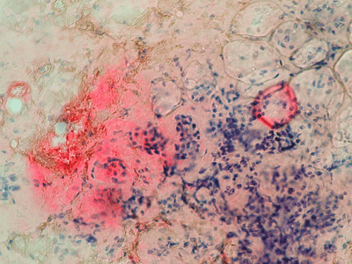
C. Congo red |
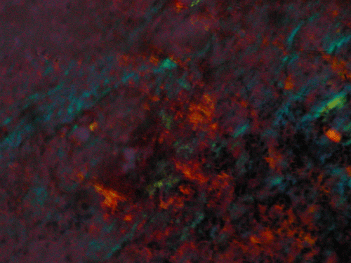
D. Congo red polarized |
E. Congo red |
F. Congo red polarized |
Pathology:
Clumps of amorphous substances with hypocellularity are noted with DiffQuick stain (arrows in Panel A) and Papanicolaou stain (arrows in Panel B). Normal glandular structures are also seen these preparations (Panel A and B). These amorphous substance stains orange on Congo red stain (Panel C and E) which gives a birefrigence under polarized light (Panel D and F are corresponding images of Panel C and E). No tumor tissue is identified.
| DIAGNOSIS: Amyloidosis of Salivary Gland. |
Discussion: General Pathogenesis Pathology Diagnosis Prognosis
General Information
Amyloidosis is a broad designation for diseases characterized by the extracellular deposition and accumulation of amyloid. Amyloid is an insoluble protein with characteristic properties. It appears as non-branching fibrils with an average diameter of 10-12 nm. At the molecular level, the protein molecules arrange in a beta-pleated sheet conformation. This beta-pleated sheet is responsible for the distinctive apple green birefringence with Congo red stain 1. With routine hematoxylin-eosin stain, amyloid appears as pale eosinophilic, amorphous, extracellular deposition 1, 2.
Amyloid is composed of protein with abnormal conformation and the source of protein is quite diversified. The AL (amyloid light chain) and AA (amyloid-associated) types are the most common. AL proteins are produced by plasma cells and is associated with plasma cell dyscrasias such as multiple myeloma. AA proteins are produced by the liver and are associated with chronic inflammation such as tuberculosis, osteomyelitis, and other chronic inflammatory conditions. Other forms of amyloid protein include transthyretin, Beta2-microglobulin, and amyloid-b protein. Amyloid can be localized to a single organ or can be systemic involving several organ systems.
Amyloid deposition can be localized to a tissue or organ or systemic. Amyloid deposition is often, but not always, associated with an underlying illness with chronic inflammatory conditions and presence of abnormal proteins in serum as the most common causes. Some tumors, such as medullary carcinoma of the thyroid gland also lead to amyloid deposition within the tumor tissue.
Primary amyloidosis is caused by the secretion of an abnormal immunoglobulin by a clonal proliferation of cells with 20% of cases present as an overt multiple myeloma. The signs and symptoms of primary systemic amyloidosis result from the deposition of light chain-containing amyloid fibrils in various organs. Patients often present with features of nephrotic syndrome or renal failure, congestive heart failure, carpal tunnel syndrome, sensorimotor peripheral neuropathy, and orthostastic hypotension. Common symptoms of primary systemic amyloidosis include weakness, fatigue, weight loss, edema, paresthesias, light-headedness or syncope, dyspnea, purpura, claudication, bleeding, or voice change. In the United States, primary systemic amyloidosis accounts for about 85% of cases of systemic amyloidosis.
Secondary amyloidosis is associated with chronic inflammatory conditions such as rheumatoid arthritis, connective tissue disorders, and inflammatory bowel disease. The depositions of the amyloid in secondary amyloidosis can be local or systemic. The distinction of the two forms is very important, since the treatment options as well as prognosis are different.
Primary amyloidosis tends to involve the heart, gastrointestinal tract, nerves, skin, tongue, eye, and respiratory system. Secondary amyloidosis tends to involve the kidneys, liver, spleen, lymph nodes, adrenals, and thyroid. Symptomatic amyloidosis of the head and neck is a rare but well documented condition 3, 4, 5, 6, 7. Most cases in this region involve one organ, without evidence of generalized involvement. The cytologic diagnosis of unexpected amyloidosis in the head and neck region can be challenging. Amyloid can be misinterpreted as keratin debris, colloid, chondroid or basement membrane material. Salivary gland amyloidosis is most often secondary and can mimic Sjogren’s syndrome and also pleomorphic adenoma because of the amorphous appearance of amyloid. Amyloidosis involving the major salivary gland in the form of a mass is rather uncommon and often present as diffuse or focal, bilateral enlargement of salivary glands. Variable amounts of acellular, eosinophilic extracellular material stains pale red with Congo red stain, and exhibits a characteristic apple-green birefringence under polarized light. The remainder of the smear is often hypocellular, with scant or absent acinar cells, and scattered clusters of ductal epithelial cells such as are seen in chronic sialadenitis.
While symptomatic manifestation of amyloid in the head and neck organs as mass or nodules are rare, labial biopsy has been used in the diagnosis of amyloidosis 8, 9. Biopsy of adipose has also been used for the diagnosis of amyloidosis 10. P-component tends to co-deposit with amyloid. 123I labeled P-component has been recently applied to detect amyloid in vivo 11, 12.
In our patient under discussion, the combination of amyloidosis with a monoclonal immunoglobin light chain suggests AL type amyloidosis. AL amyloidosis is more often due to monoclonal lambda light chains than kappa light chains. The bone marrow in this case showed a slight increase in plasma cells, some of which appeared cytologically atypical, suggesting a plasma cell dyscrasia.
While the localized forms of amyloidosis have relatively good prognosis and can be managed by excision or conservative treatment, the systemic forms require treatment of underlying conditions and usually have a poor prognosis. Although amyloidosis is a multisystemic disease, the prognosis is determined primarily by the presence or absence of cardiac involvement. Patients with overt congestive heart failure survive from 4 to 6 months and is the single most important prognostic factor. Patients who do not have congestive heart failure have a median survival of 30 months. Determination of whether cardiac involvement is present is of the utmost importance in counseling the patient. Two-dimensional Doppler echocardiography, to study diastolic filling, is a critical technique for recognizing the presence or absence of amyloid. The ejection fraction is an important predictor of survival in a patient with amyloidosis. The thickness of the interventricular septum in diastole also is predictive of survival because it seems to reflect the amount of amyloid deposited in the myocardium. Doppler echocardiographic studies of left ventricular filling reflect the degree of stiffness of the heart and can indicate the presence of restrictive hemodynamics. Echocardiography is considered a standard test for all patients with a diagnosis of amyloidosis, without regard to the initial site of involvement.
Reference:
Cotran R, Kumar V, Robbins S. Amyloidosis. Pathologic Basis of Disease. WB Saunders Co; 1999:231-238.
Gertz MA, Lacy MQ, Dispenzieri A. Amyloidosis: Recognition, Confirmation, Prognosis, and Therapy. Mayo Clinic Proceedings. 1999 74:490-494.
Kolokotronis A, Stefanopoulos P, Xohellis M, Antoniadas D. Oral Manifestations of Amyloidosis: Report of Two Cases. Oral Surgery, Oral Medicine, Oral Pathology, Oral Radiology, and Endodontics. 2004;98:200-201.
Giorgadze T, Baloch Z, Thaler E, Gupta P. Unsuspected Systemic Amyloidosis Diagnosed by Fine-Needle Aspiration of the Salivary Gland: Case Report. Diagnostic Cytopathology. 2004 31:57-59.
Koloktronis A, Chatzigiannis I, Paloukidou N. Oral Involvement in a Case of AA Amyloidosis. Oral Dis. 2003 9:269-72.
Mateo Arias J, Molina Martinez M, Borrego A, Mayorga F. Amyloidosis of the submaxillary gland. Med Oral. 2003 8:66-70.
Sbai A, Wechsler B, Leenhardt L, Beaufils H, Hoang C, Menegaux F, Piette JC. Amyloid goiter as the initial manifestation of systemic amyloidosis due to familial mediterranean fever with homozygous MEFV mutation. Thyroid. 2001 11:397-400.
Hachulla E, Janin A, Flipo RM, Saile R, Facon T, Bataille D, Vanhille P, Hatron PY, Devulder B, Duquesnoy B. Labial salivary gland biopsy is a reliable test for the diagnosis of primary and secondary amyloidosis. A prospective clinical and immunohistologic study in 59 patients. Arthritis Rheum. 1993 36:691-7.
Delgado WA, Mosqueda A. A highly sensitive method for diagnosis of secondary amyloidosis by labial salivary gland biopsy. J Oral Pathol Med. 1989 18:310-4.
Andrews TR, Colon-Otero G, Calamia KT, Menke DM, Boylan KB, Kyle RA. Utility of subcutaneous fat aspiration for diagnosing amyloidosis in patients with isolated peripheral neuropathy. Mayo Clin Proc. 2002 77:1287-1290.
Hazenberg BP, van Rijswijk MH, Piers DA, Lub-de Hooge MN, Vellenga E, Haagsma EB, Hawkins PN, Jager PL. Diagnostic performance of 123I-labeled serum amyloid P component scintigraphy in patients with amyloidosis. Am J Med. 2006 119:355.e15-24.
Maulin L, Hachulla E, Deveaux M, Janin A, Wechsler B, Godeau P, Rousset H, Barrier JH, Hatron PY, Devulder B, Huglo D, Marchandise X. 'Localized amyloidosis': 123I-labelled SAP component scintigraphy and labial salivary gland biopsy. QJM. 1997 90:45-50.
Cases of the Month Evaluation Coordinator: KarMing-Fung@ouhsc.edu